Review
. 2011:73:213-37.
doi: 10.1146/annurev.physiol.70.113006.100646.

Mesenchymal cells of the intestinal lamina propria

Affiliations
Review

Mesenchymal cells of the intestinal lamina propria

D W Powell et al. Annu Rev Physiol. 2011.

Abstract

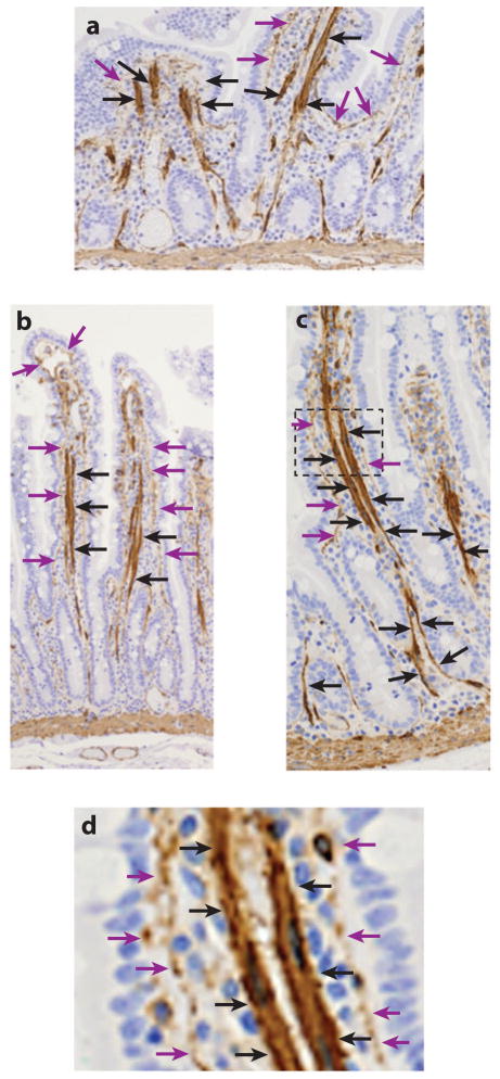
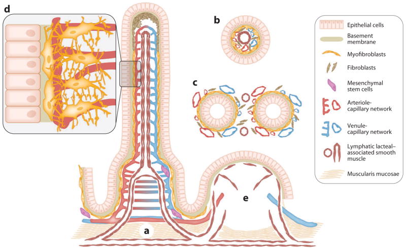
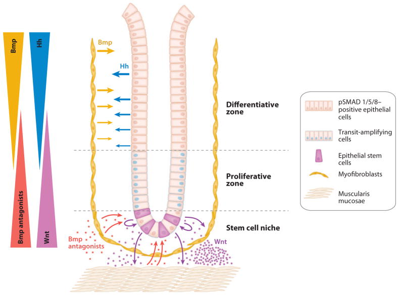
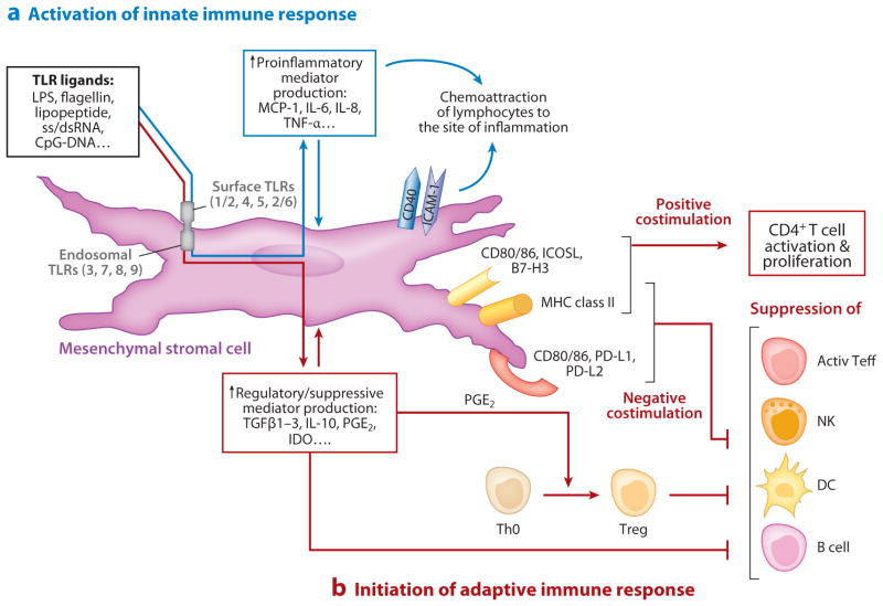
The mesenchymal elements of the intestinal lamina propria reviewed here are the myofibroblasts, fibroblasts, mural cells (pericytes) of the vasculature, bone marrow-derived stromal stem cells, smooth muscle of the muscularis mucosae, and smooth muscle surrounding the lymphatic lacteals. These cells share similar marker molecules, origins, and coordinated biological functions previously ascribed solely to subepithelial myofibroblasts. We review the functional anatomy of intestinal mesenchymal cells and describe what is known about their origin in the embryo and their replacement in adults. As part of their putative role in intestinal mucosal morphogenesis, we consider the intestinal stem cell niche. Lastly, we review emerging information about myofibroblasts as nonprofessional immune cells that may be important as an alarm system for the gut and as a participant in peripheral immune tolerance.


Figures

Figure 1
Figure 1
α-Smooth muscle actin (α-SMA) immunohistology (brown) of the human jejunum. (a) Longitudinal section through a poorly oriented jejunum shows what appears to be random smooth muscle fibers oriented in a longitudinal fashion in villus core. (b,c) Better-oriented sections of (b) the upper aspects and (c) the lower aspects of the villus-crypt axis. In these sections the smooth muscle bundles are closely associated with the lymphatic lacteals, which are covered in their lower aspects after they bifurcate to enter the muscularis by only pericytes rather than organized smooth muscle bundles (black arrows). α-SMA+ subepithelial myofibroblasts and vascular mural cells (pericytes) are located between the epithelium and the lacteal (purple arrows). (d ) An enlarged section of panel c shows that the lacteal and the subepithelial myofibroblasts are separate α-SMA+ elements. Figure courtesy of Patrick Adegboyega, Louisiana State University Health Sciences Center, Shreveport, Louisiana.
Figure 2
Figure 2
The spatial relationships of the epithelium and mesenchymal elements of the small intestinal villus-crypt axis. (a) Longitudinal representation of the villus and crypt showing the epithelium and lamina propria containing α-SMA+ subepithelial myofibroblasts and pericytes of the capillaries, mesenchymal stem cells, and smooth muscle associated with the lymphatic lacteal and the muscularis mucosae. Fibroblasts (α-SMA) are shown, especially in the upper portion of the villus. (b) Cross sections through a villus show the lymphatic lacteal with associated smooth muscle. (c) A cross section through the crypts demonstrates the lymphatic pericytes. Panels b and c also show that the subepithelial myofibroblasts are essentially pericytes in subepithelial locations of the villus. (d ) A higher-power depiction of the myofibroblasts and pericytes with cytoplasmic processes that surround and support the capillaries. (e) A Peyer’s patch with its vascular, lymphatic, and stromal elements. Lymphocytes, macrophages, dendritic cells, and polymorphonuclear leukocytes are not shown.
Figure 3
Figure 3
Epithelial-mesenchymal interactions orchestrate hedgehog (Hh), bone morphogenetic protein (Bmp), and Wnt signaling in the intestinal crypt. Hh proteins produced by differentiated epithelial cells trigger stromal Bmp synthesis. Active Bmp signaling within differentiated epithelial cells, evidenced by phosphorylated SMADs (pSMAD) 1, 5, and 8, contributes to maintenance of the differentiated state and proliferative quiescence. Within the stem cell niche, lack of Hh signaling leads to decreased stromal Bmp synthesis. Secretion of canonical Wnts by crypt-based epithelial and Paneth cells, in addition to promoting self-renewal of intestinal epithelial stem cells, may also induce synthesis and secretion of Bmp antagonists by the underlying myofibroblasts and muscularis mucosae cells (85). Bmp antagonism within the stem cell niche would further enhance Wnt signaling. Not shown in this model are autocrine effects of Bmp signaling upon the stromal cells or numerous other factors, stromal and epithelial, that regulate differentiation and self-renewal within intestinal crypts (64). Adapted from Kosinski et al. (84), with permission.
Figure 4
Figure 4
Mesenchymal stromal cells as links between innate and adaptive immunity. (a) Stromal cells participate in innate immunity via expression of TLRs, whose engagement induces expression of either pro- or anti-inflammatory soluble mediators and surface-associated molecules. The proinflammatory molecules (e.g., MCP-1, IL-8) induce migration of professional immune cells where they may be anchored via interaction with stromal adhesive molecules (e.g., ICAM-1, CD40). (b) Adaptive immunity is initiated via signaling through MHC class II molecules and B7-positive or B7-negative costimulators expressed on mesenchymal stromal cells modulating the activity of CD4+ T cells, NK, DC, and B cells. Depending on the microenvironmental milieu of cytokines, chemokines, growth factors, and pathogen-associated molecular patterns (PAMPs), stromal cells function in either an immunosuppressive (tolerogenic) role or an immunostimulatory role. Abbreviations: active Teff, activated effector T cells; CD, cluster of differentiation; ICAM-1, intercellular adhesion molecule-1; IDO, indoleamine 2,3-dioxygenase; IL, interleukin; NK, natural killer cells; DC, dendritic cells; MCP-1, monocyte chemotactic protein-1; MHC, major histocompatibility complex; PD-L, programmed cell death receptor ligand; PGE2, prostaglandin E2; ss/dsRNA, single-stranded/double-stranded RNA; TGFβ, transforming growth factor β; Th0, naive CD4+ T helper cells; TLR, Toll-like receptor; TNF-α, tumor necrosis factor-α; Treg, CD4+ CD25high FoxP3+ regulatory T cells.

References

LITERATURE CITED

    1. Powell DW, Adegboyega PA, Di Mari JF, Mifflin RC. Epithelial cells and their neighbors. I Role of intestinal myofibroblasts in development, repair, and cancer. Am J Physiol Gastrointest Liver Physiol. 2005;289:2–7. - PubMed
    1. Powell DW, Mifflin RC, Valentich JD, Crowe SE, Saada JI, West AB. Myofibroblasts. II Intestinal subepithelial myofibroblasts. Am J Physiol Cell Physiol. 1999;277:183–201. - PubMed
    1. Powell DW, Mifflin RC, Valentich JD, Crowe SE, Saada JI, West AB. Myofibroblasts. I Paracrine cells important in health and disease. Am J Physiol Cell Physiol. 1999;277:1–9. - PubMed
    1. Hinz B, Phan SH, Thannickal VJ, Galli A, Bochaton-Piallat ML, Gabbiani G. The myofibroblast: one function, multiple origins. Am J Pathol. 2007;170:1807–16. - PMC - PubMed
    1. Simon-Assmann P, Bolcato-Bellemin AL, Klein A, Kedinger M. Tissue recombinants to study extracellular matrix targeting to basement membranes. Methods Mol Biol. 2009;522:309–18. - PubMed

RELATED RESOURCES

    1. Kalluri R, Zeisberg M. Fibroblasts in cancer. Nat Rev Cancer. 2006;6:392–401. - PubMed
    1. Roberts DJ. Molecular mechanisms of development of the gastrointestinal tract. Dev Dyn. 2000;219:109–20. - PubMed
    1. McLin VA, Henning SJ, Jamrich M. The role of the visceral mesoderm in the development of the gastrointestinal tract. Gastroenterology. 2009;136:2074–91. - PubMed
    1. Rubin DC. Intestinal morphogenesis. Curr Opin Gastroenterology. 2007;23:111–14. - PubMed
    1. Mueller SN, Germain RN. Stromal cell contributions to the homeostasis and functionality of the immune system. Nat Rev Immunol. 2009;9:618–29. - PMC - PubMed

Publication types

MeSH terms