

Identification of telocytes in the lamina propria of rat duodenum: transmission electron microscopy
- PMID: 21054782
- PMCID: PMC3822490
- DOI: 10.1111/j.1582-4934.2010.01207.x
Identification of telocytes in the lamina propria of rat duodenum: transmission electron microscopy
Abstract
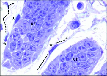
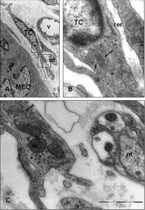
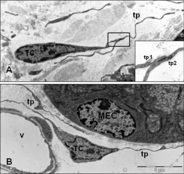
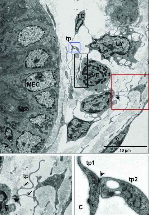
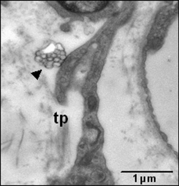
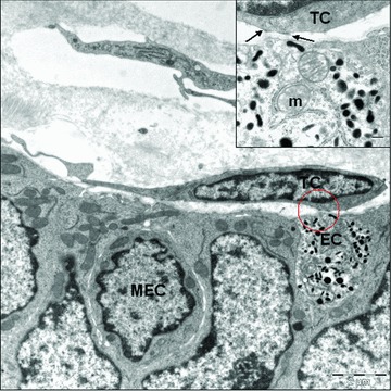
Recently the new term 'telocytes' has been proposed for cells formerly known as interstitial Cajal-like cells. In fact, telocytes are not really Cajal-like cells, they being different from all other interstitial cells by the presence of telopodes, which are cell-body prolongations, very thin, extremely long with a moniliform aspect. The identification of these cells is based on ultrastructural criteria. The presence of telocytes in others organs was previously documented. We reported for the first time, an ultrastructural study of telocytes in the lamina propria of rat duodenum. Our findings show that typical telocytes are present in the rat duodenum. Telocytes are located in the lamina propria, immediately below mucosal crypts. Telopodes frequently establish close spatial relationships with immune cells, blood vessels and nerve endings. On the basis of their distribution and morphology, we suggest that these cells may be involved in immune response and in our opinion, it may be possible that different locations of telocytes could be associated with different roles.
© 2011 The Authors Journal of Cellular and Molecular Medicine © 2011 Foundation for Cellular and Molecular Medicine/Blackwell Publishing Ltd.
Figures
Similar articles
-
Telocytes in endocardium: electron microscope evidence.J Cell Mol Med. 2010 Sep;14(9):2330-4. doi: 10.1111/j.1582-4934.2010.01133.x. J Cell Mol Med. 2010. PMID: 20716125 Free PMC article.
-
Cellular Evidence of Telocytes as Novel Interstitial Cells Within the Magnum of Chicken Oviduct.Cell Transplant. 2017 Jan 24;26(1):135-143. doi: 10.3727/096368916X692942. Epub 2016 Sep 1. Cell Transplant. 2017. PMID: 27590447 Free PMC article.
-
Telocytes, a distinct type of cell among the stromal cells present in the lamina propria of jejunum.Histol Histopathol. 2012 Aug;27(8):1067-78. doi: 10.14670/HH-27.1067. Histol Histopathol. 2012. PMID: 22763879
-
Myocardial telocytes: a specific new cellular entity.J Cell Mol Med. 2010 Jul;14(7):1917-21. doi: 10.1111/j.1582-4934.2010.01111.x. Epub 2010 Jul 5. J Cell Mol Med. 2010. PMID: 20604817 Free PMC article. Review.
-
Telocytes and interstitial cells of Cajal in the biliary system.J Cell Mol Med. 2018 Jul;22(7):3323-3329. doi: 10.1111/jcmm.13643. Epub 2018 Apr 26. J Cell Mol Med. 2018. PMID: 29700981 Free PMC article. Review.
Cited by
-
Altered distribution of interstitial cells and innervation in the rat urinary bladder following spinal cord injury.J Cell Mol Med. 2012 Jul;16(7):1533-43. doi: 10.1111/j.1582-4934.2011.01410.x. J Cell Mol Med. 2012. PMID: 21883887 Free PMC article.
-
Cardiac telocytes exist in the adult Xenopus tropicalis heart.J Cell Mol Med. 2020 Feb;24(4):2531-2541. doi: 10.1111/jcmm.14947. Epub 2020 Jan 12. J Cell Mol Med. 2020. PMID: 31930692 Free PMC article.
-
Telocytes and stem cells in limbus and uvea of mouse eye.J Cell Mol Med. 2013 Aug;17(8):1016-24. doi: 10.1111/jcmm.12111. J Cell Mol Med. 2013. PMID: 23991685 Free PMC article.
-
Simulation and Modeling of Telocytes Behavior in Signaling and Intercellular Communication Processes.Int J Mol Sci. 2020 Apr 9;21(7):2615. doi: 10.3390/ijms21072615. Int J Mol Sci. 2020. PMID: 32283771 Free PMC article.
-
Telocytes: ultrastructural, immunohistochemical and electrophysiological characteristics in human myometrium.Reproduction. 2013 Apr 15;145(4):357-70. doi: 10.1530/REP-12-0369. Print 2013 Apr. Reproduction. 2013. PMID: 23404846 Free PMC article.
References
-
- Ramón y, Cajal S. Notes on the Auerbach’s plexus of the frog. Histological laboratory works of the Medicine Faculty of Barcelona. 1892:23–8.
-
- Ramón y, Cajal S. Ganglia and nerve plexus of the mammal gut and some contributions to our studies on the spine and sympathetic nervous system. Madrid: Imprenta y Librerí de Nicolás Moya. 1893:5–37.
-
- Davidson RA, McCloskey KD. Morphology and localization of interstitial cells in the guinea pig bladder: structural relationships with smooth muscle and neurons. J Urol. 2005;173:1385–90. - PubMed
Publication types
MeSH terms
LinkOut - more resources
Full Text Sources
