

Arginase II deletion increases corpora cavernosa relaxation in diabetic mice
- PMID: 21054801
- PMCID: PMC3117078
- DOI: 10.1111/j.1743-6109.2010.02098.x
Arginase II deletion increases corpora cavernosa relaxation in diabetic mice
Abstract
Introduction: Diabetes-induced erectile dysfunction involves elevated arginase (Arg) activity and expression. Because nitric oxide (NO) synthase and Arg share and compete for their substrate L-arginine, NO production is likely linked to regulation of Arg. Arg is highly expressed and implicated in erectile dysfunction.
Aim: It was hypothesized that Arg-II isoform deletion enhances relaxation function of corpora cavernosal (CC) smooth muscle in a streptozotocin (STZ) diabetic model.
Methods: Eight weeks after STZ-induced diabetes, vascular functional studies, Arg activity assay, and protein expression levels of Arg and constitutive NOS (using Western blots) were assessed in CC tissues from nondiabetic wild type (WT), diabetic (D) WT (WT + D), Arg-II knockout (KO), and Arg-II KO+D mice (N = 8-10 per group).
Main outcome measures: Inhibition or lack of arginase results in facilitation of CC relaxation in diabetic CC.
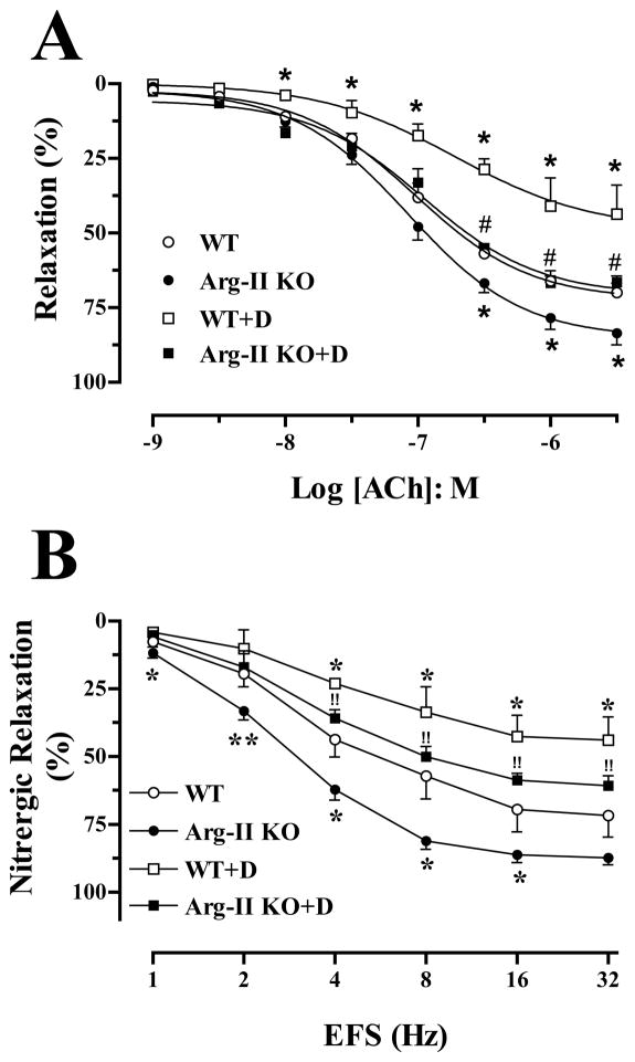
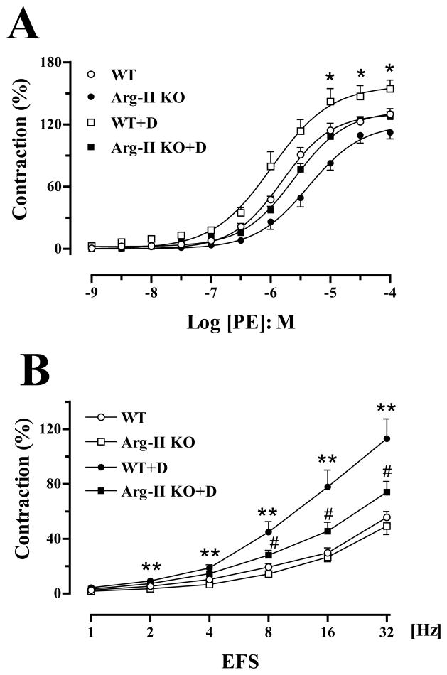
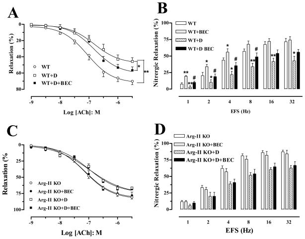
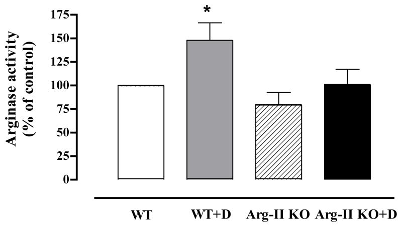
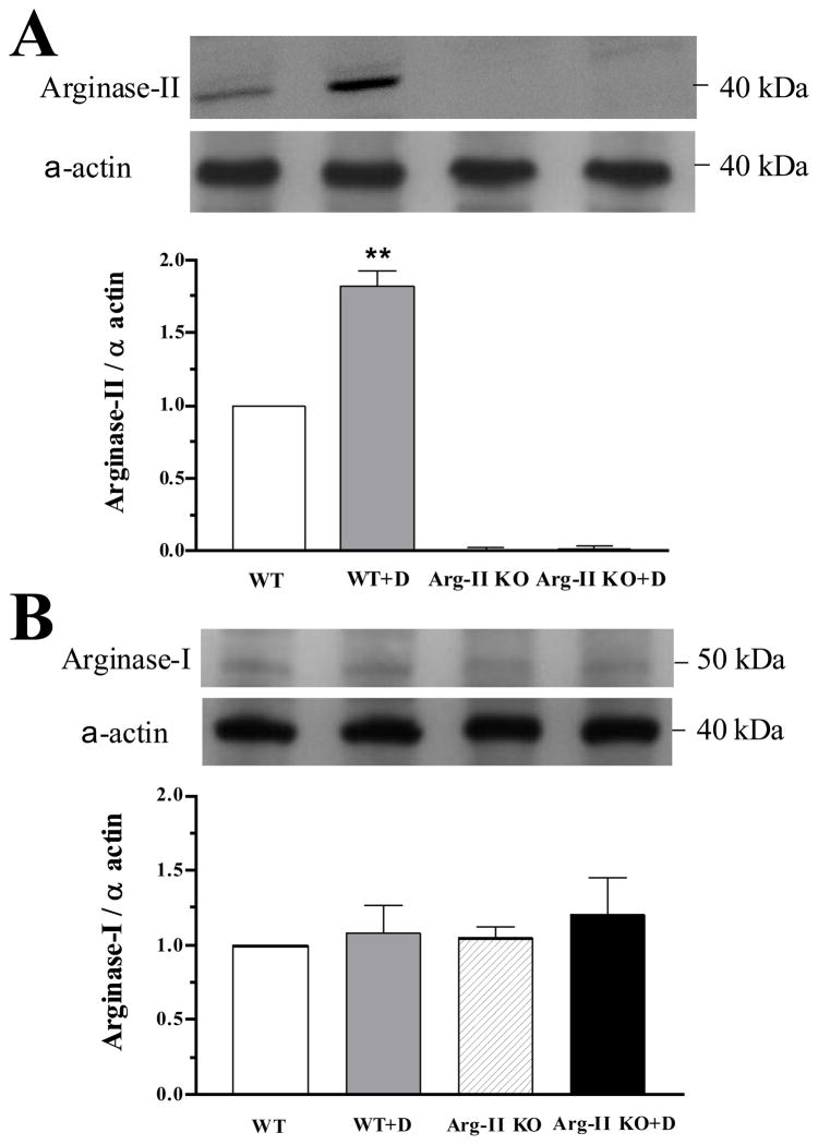
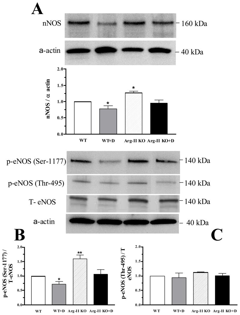
Results: Strips of CC from Arg-II KO mice exhibited an enhanced maximum endothelium-dependent relaxation (from 70 + 3% to 84 + 4%) and increased nitrergic relaxation (by 55%, 71%, 42%, 42%, and 24% for 1, 2, 4, 8 and 16 Hz, respectively) compared with WT mice. WT + D mice showed a significant reduction of endothelium-dependent maximum relaxation (44 + 8%), but this impairment of relaxation was significantly prevented in Arg-II KO+D mice (69 + 4%). Sympathetic-mediated and alpha-adrenergic agent-induced contractile responses also were increased in CC strips from D compared with non-D controls. Contractile responses were significantly lower in Arg-II KO control and D versus the WT groups. WT + D mice increased Arg activity (1.5-fold) and Arg-II protein expression and decreased total and phospho-eNOS at Ser-1177, and nNOS levels. These alterations were not seen in Arg-II KO mice. Additionally, the Arg inhibitor BEC (50 µM) enhanced nitrergic and endothelium-dependent relaxation in CC of WT + D mice.
Conclusion: These studies show for the first time that Arg-II deletion improves CC relaxation in type 1 diabetes.
© 2010 International Society for Sexual Medicine.
Conflict of interest statement
Conflict of Interest: None
Figures
Similar articles
-
Akita spontaneously type 1 diabetic mice exhibit elevated vascular arginase and impaired vascular endothelial and nitrergic function.PLoS One. 2013 Aug 19;8(8):e72277. doi: 10.1371/journal.pone.0072277. eCollection 2013. PLoS One. 2013. PMID: 23977269 Free PMC article.
-
Activated Rho kinase mediates diabetes-induced elevation of vascular arginase activation and contributes to impaired corpora cavernosa relaxation: possible involvement of p38 MAPK activation.J Sex Med. 2013 Jun;10(6):1502-15. doi: 10.1111/jsm.12134. Epub 2013 Apr 8. J Sex Med. 2013. PMID: 23566117 Free PMC article.
-
Extracellular signal-regulated kinase (ERK) inhibition decreases arginase activity and improves corpora cavernosal relaxation in streptozotocin (STZ)-induced diabetic mice.J Sex Med. 2011 Dec;8(12):3335-44. doi: 10.1111/j.1743-6109.2011.02499.x. Epub 2011 Oct 13. J Sex Med. 2011. PMID: 21995824
-
p38 Mitogen-activated protein kinase (MAPK) increases arginase activity and contributes to endothelial dysfunction in corpora cavernosa from angiotensin-II-treated mice.J Sex Med. 2010 Dec;7(12):3857-67. doi: 10.1111/j.1743-6109.2010.01996.x. Epub 2010 Aug 30. J Sex Med. 2010. PMID: 20807329 Free PMC article.
-
TNF-alpha knockout mice have increased corpora cavernosa relaxation.J Sex Med. 2009 Jan;6(1):115-25. doi: 10.1111/j.1743-6109.2008.01029.x. J Sex Med. 2009. PMID: 19170842 Free PMC article.
Cited by
-
Arginase promotes skeletal muscle arteriolar endothelial dysfunction in diabetic rats.Front Immunol. 2013 May 20;4:119. doi: 10.3389/fimmu.2013.00119. eCollection 2013. Front Immunol. 2013. PMID: 23730303 Free PMC article.
-
Role of Metabolites of Nitric Oxide and Arginase in the Pathogenesis of Glomerulonephritis.Curr Health Sci J. 2016 Jul-Sep;42(3):221-225. doi: 10.12865/CHSJ.42.03.01. Epub 2016 Sep 29. Curr Health Sci J. 2016. PMID: 30581575 Free PMC article.
-
Deficient arginase II expression without alteration in arginase I expression attenuated experimental autoimmune encephalomyelitis in mice.Immunology. 2018 Sep;155(1):85-98. doi: 10.1111/imm.12926. Epub 2018 Apr 16. Immunology. 2018. PMID: 29574762 Free PMC article.
-
Akita spontaneously type 1 diabetic mice exhibit elevated vascular arginase and impaired vascular endothelial and nitrergic function.PLoS One. 2013 Aug 19;8(8):e72277. doi: 10.1371/journal.pone.0072277. eCollection 2013. PLoS One. 2013. PMID: 23977269 Free PMC article.
-
Angiotensin II-induced arterial thickening, fibrosis and stiffening involves elevated arginase function.PLoS One. 2015 Mar 25;10(3):e0121727. doi: 10.1371/journal.pone.0121727. eCollection 2015. PLoS One. 2015. PMID: 25807386 Free PMC article.
References
-
- Andersson KE. Pharmacology of penile erection. Pharmacol Rev. 2001;53:417–50. - PubMed
-
- Toque HA, Romero MJ, Tostes RC, Shatanawi A, Chandra S, Carneiro Z, Inscho E, Webb RC, Caldwell RB, Caldwell RW. p38 Mitogen-Activated Protein Kinase (MAPK) Increases Arginase Activity and Contributes to Endothelial Dysfunction in Corpora Cavernosa from Angiotensin-II Treated Mice. JSM. 2010 In Press. - PMC - PubMed
-
- Xie D, Odronic SI, Wu F, Pippen AM, Donatucci CF, Annex BH. A mouse model of hypercholesterolemia-induced erectile dysfunction. J Sex Med. 2007;4:898–907. - PubMed
-
- Bivalacqua TJ, Burnett AL, Hellstrom WJ, Champion HC. Overexpression of arginase in the aged mouse penis impairs erectile function and decreases eNOS activity: influence of in vivo gene therapy of anti-arginase. Am J Physiol Heart Circ Physiol. 2007;292:H1340–51. - PubMed
-
- Bivalacqua TJ, Hellstrom WJ, Kadowitz PJ, Champion HC. Increased expression of arginase II in human diabetic corpus cavernosum: in diabetic-associated erectile dysfunction. Biochem Biophys Res Commun. 2001;283:923–7. - PubMed
Publication types
MeSH terms
Substances
Grants and funding
LinkOut - more resources
Full Text Sources
Research Materials
