. 2010 Nov 5:7:93.
doi: 10.1186/1742-4690-7-93.

Misfolding of CasBrE SU is reversed by interactions with 4070A Env: implications for gammaretroviral neuropathogenesis

Affiliations

Misfolding of CasBrE SU is reversed by interactions with 4070A Env: implications for gammaretroviral neuropathogenesis

Ying Li et al. Retrovirology. .

Abstract

Background: CasBrE is a neurovirulent murine leukemia virus (MLV) capable of inducing paralytic disease with associated spongiform neurodegeneration. The neurovirulence of this virus has been genetically mapped to the surface expressed subunit (SU) of the env gene. However, CasBrE SU synthesized in the absence of the transmembrane subunit (TM) does not retain ecotropic receptor binding activity, indicating that folding of the receptor binding domain (RBD) requires this domain. Using a neural stem cell (NSC) based viral trans complementation approach to examine whether misfolded CasBrE SU retained neurovirulence, we observed CasBrE SU interaction with the "non-neurovirulent" amphotropic helper virus, 4070A which restored functional activity of CasBrE SU.

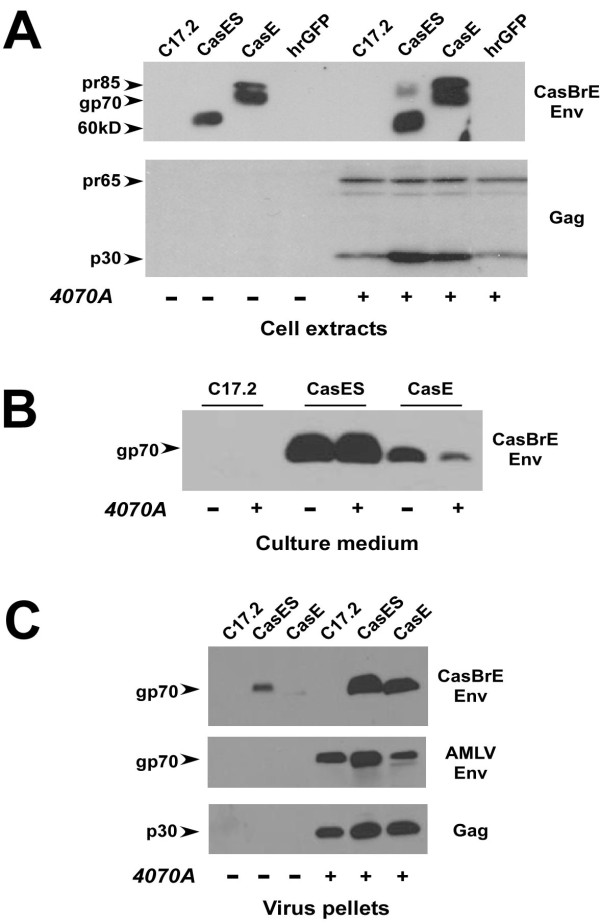
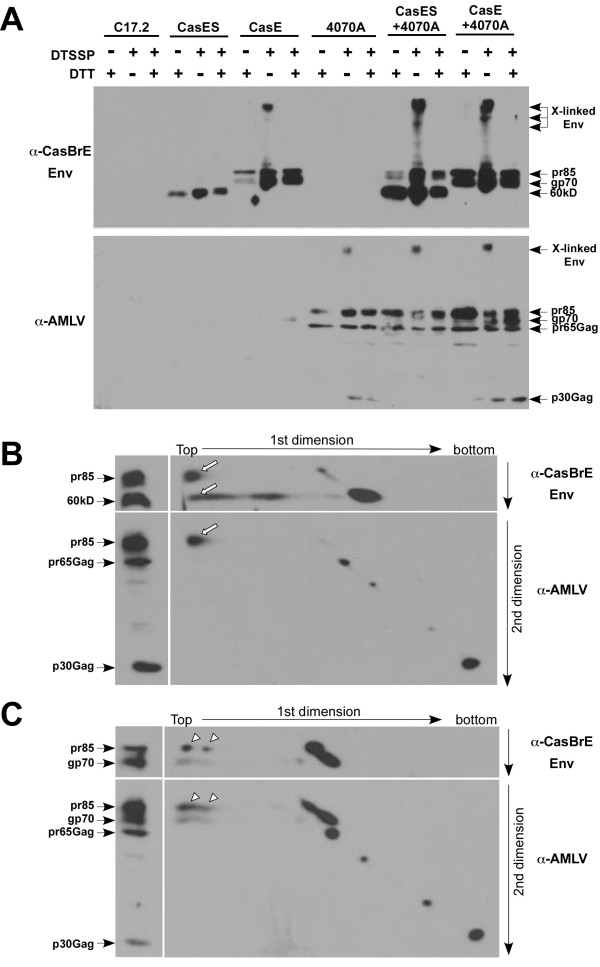
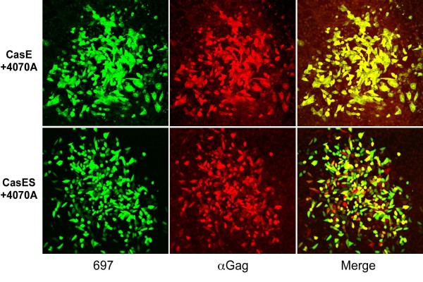
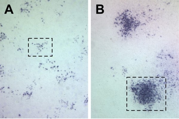
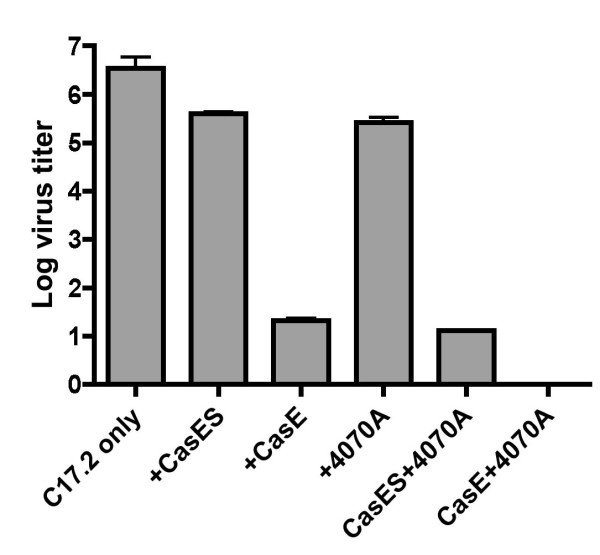
Results: Herein, we show that infection of NSCs expressing CasBrE SU with 4070A (CasES+4070A-NSCs) resulted in the redistribution of CasBrE SU from a strictly secreted product to include retention on the plasma membrane. Cell surface cross-linking analysis suggested that CasBrE SU membrane localization was due to interactions with 4070A Env. Viral particles produced from CasES+4070A-NSCS contained both CasBrE and 4070A gp70 Env proteins. These particles displayed ecotropic receptor-mediated infection, but were still 100-fold less efficient than CasE+4070A-NSC virus. Infectious center analysis showed CasBrE SU ecotropic transduction efficiencies approaching those of NSCs expressing full length CasBrE Env (CasE; SU+TM). In addition, CasBrE SU-4070A Env interactions resulted in robust ecotropic superinfection interference indicating near native intracellular SU interaction with its receptor, mCAT-1.

Conclusions: In this report we provided evidence that 4070A Env and CasBrE SU physically interact within NSCs leading to CasBrE SU retention on the plasma membrane, incorporation into viral particles, restoration of mCAT-1 binding, and capacity for initiation of TM-mediated fusion events. Thus, heterotropic Env-SU interactions facilitates CasBrE SU folding events that restore Env activity. These findings are consistent with the idea that one protein conformation acts as a folding scaffold or nucleus for a second protein of similar primary structure, a process reminiscent of prion formation. The implication is that template-based protein folding may represent an inherent feature of neuropathogenic proteins that extends to retroviral Envs.


Figures

Figure 1
Figure 1
Structures of the amphotropic virus and retroviral vectors introduced into NSCs for pseudotyping and trans complementation analysis of CasBrE env genes. C17.2 NSCs were transduced with the pSFF-based retroviral vectors encoding CasBrE SU/TM (CasE), CasBrE SU (CasES), or the humanized renilla green fluorescent protein (hrGFP; striped) followed by infection with the amphotropic virus 4070A. The complementing viral structural elements are shown as filled regions for the 4070A virus and CasE and CasES vectors. The hrGFP vector served as a vector control that could be followed by fluorescence microscopy. Vector elements designated with a Δ represent deleted retroviral structural elements. Ψ indicates the presence of retroviral packaging sequences. SD, Splice donor. SA, Splice acceptor. LTR, Long terminal repeat.
Figure 2
Figure 2
FACS analysis of control, CasE, CasES, and hrGFP-vector transduced C17.2 NSCs, with and without 4070A infection. The FACS analysis of CasES- and CasE-NSCs shown was limited to unpermeabilized cells in order to show cell surface CasBrE Env expression. CasBrE Env expression levels were detected via staining with the monoclonal antibody designated 697 [32]. GFP expression was detected directly.
Figure 3
Figure 3
Western blotting analysis of vector transduced NSCs without (-) and with (+) 4070A infection. A. Equivalent whole cell extracts of control C17.2 NSCs and those expressing CasE, CasES or hrGFP, with and without 4070A infection, were assessed for CasBrE Env expression (αCasBrE Env; top) and Gag (αGag; bottom) after separation on 9% SDS-PAGE gels. CasES-NSCs produce a 60 kD Env isoform in contrast to the gp70 and precursor (pr85) isoforms made in CasE-NSCs [11]. The 60 kD isoform likely represents minimally glycosylated Env protein [40]. Note that CasBrE Env levels were elevated with 4070A infection, and p30gag levels are elevated in 4070A infected NSCs possessing CasBrE Env vectors. B. Examination of CasBrE Env released into the culture medium ± 4070A infection. Note that CasBrE gp70 release is reduced in the presence of 4070A infection for CasE-NSCs but not CasES NSCs. C. NSC culture supernatants were subjected to ultracentrifugation and virion pellets were assessed for CasBrE Env (top), 4070A Env (middle) and Gag (bottom). All blots were run in triplicate using equivalent protein loads that were confirmed by coomassie blue staining of equivalent gels run in parallel. Representative examples are shown.
Figure 4
Figure 4
Cell surface cross-linking reveals direct CasBrE Env and 4070A interactions in pseudotyping NSCs. A. Treatment of control, 4070A virus, or vector transduced C17.2 NSCs with the water soluble chemical cross-linker DTSSP to assess cell surface interactions between CasBrE SU or SU/TM and 4070A Env. Extracts from cross-linked cells were treated with or without dithiotheitol (DTT), separated by SDS-PAGE on 8% gels, followed by immunoblotting first for CasBrE Env (A-C, top panels; 697) followed by stripping and reprobing for 4070A viral proteins (A-C, lower panels; pig anti-AmLV). Cross-linked complexes (X-linked Env) appear at the top of the blots as slow mobility bands or smears, which were converted by DTT treatment to molecular mobility patterns consistent with un-crosslinked samples. B. Sequential immunoblotting of cross-linked samples from CasES+4070A-NSCs separated by non-reducing SDS-PAGE (without DTT) in the first dimension (horizontal) followed by reducing SDS-PAGE (with DTT) in the second dimension (vertical). The same sample was also treated with DTT and only run in the second dimension indicated by the separate lane (left columns) to mark the migration of the different immunoreactive components. C. Non-reducing/reducing two-dimensional SDS-PAGE analysis of crosslinked CasE+4070A NSC extracts. Arrows and arrowheads indicate the presence of the Env species that co-migrated in the high molecular weight cross-linked complexes.
Figure 5
Figure 5
Viral pseudotyping from 4070A-infected NSCs. Virus titration assays (VTAs) were performed on dunni fibroblasts using culture supernatants derived from 4070A infected CasE-, CasES-, hrGFP- and control-C17.2 NSCs for total virus (white bars) and vector only encoding virus (black bars). ND = not detected. Total virus was detected with an Env specific antibody, 83A25 that recognizes both 4070A and CasBrE envelope proteins. CasBrE Env encoding viruses were detected by monoclonal antibody 697 [32]. HrGFP encoding virus was determined by direct fluorescence detection of infected cells.
Figure 6
Figure 6
CasBrE SU mediates virus entry via the ecotropic receptor. Viral supernatants taken from 4070A-infected hrGFP-, CasES-, and CasE-NSCs were titered on NIH 3T3 fibroblasts cells with (black bars) and without (white bars) 4070A infection. The 4070A infection acts as a means to interfere with virus entry via the amphotropic receptor, PiT-2. CasBrE Env+ foci were detected using monoclonal antibody 697 and hrGFP foci were detected by direct viewing of target cell fluorescence. Assays were run in triplicate and error bars represent the standard deviation of the log of the vector titers.
Figure 7
Figure 7
Target cells are dually infected by 4070A and pseudotyped viral vectors. Infectious centers (ICs) were generated by seeding 4070A-infected CasE- and CasES-NSCs with target dunni fibroblasts followed by double immunostaining for CasBrE Env (697; red) to indicate vector infection and Gag (αGag, green) to depict 4070A virus infection. Abundant colocalization in the target fibroblasts is shown in the merged images (Merge) indicating that both the viral vector and 4070A genes were efficiently delivered to target cells. Bar = 100 μm.
Figure 8
Figure 8
Foci morphology of infectious virus derived from pseudotyping NSCs. A. Representative foci (boxed areas) from cells infected with supernatants from CasE+4070A-NSCs stained for CasBrE Env showing non-contiguous foci morphologies indicative of defective replication incompetent virus. No contiguous 697 foci were noted under conditions where single-hit infection predominated, indicating that replication competent recombinant CasBrE viruses did not readily arise from CasE+4070A-NSCs or CasES+4070A-NSCs. B. Representative foci from fibroblasts infected with CasE+4070A supernatants stained with monoclonal antibody 83A25 which recognizes both 4070A and CasBrE Envs. This image illustrates both non-contiguous and contiguous foci morphologies, indicative of defective and replication competent viruses, respectively.
Figure 9
Figure 9
Assessment of CasBrE Env binding to mCAT-1 by superinfection interference. Control, CasES- and CasE-C17.2 NSCs with and without 4070A infection were subjected to infection by the ecotropic virus, Fr57E, to assess the ability of NSCs-expressing CasBrE Env or SU to block Fr57E entry through the ecotropic virus receptor mCAT-1. Viral foci on NSCs were detected with monoclonal antibodies 48, 500, and 720 that are specific for the Friend virus Env [38,39]. Error bars represent the standard deviation of the log of the Fr57E virus titers. N = 4 for all the samples.

Similar articles

Cited by

References

    1. Gardner MB. Retroviral spongiform polioencephalomyelopathy. Rev Infect Dis. 1985;7:99–110. - PubMed
    1. Gardner MB, Henderson BE, Officer JE, Rongey RW, Parker JC, Oliver C, Estes JD, Huebner RJ. A spontaneous lower motor neuron disease apparently caused by indigenous type-C RNA virus in wild mice. JNCI. 1973;51:1243–1254. - PMC - PubMed
    1. Gardner MB, Rasheed S, Klement V, Rongey RW, Brown JC, Dworsky R, Henderson BE. Lower motor neuron disease in wild mice caused by indigenous type C virus and search for a similar etiology in human amyotrophic lateral sclerosis. UCLA Forum Med Sci. 1976;19:217–234. - PubMed
    1. DesGroseillers L, Barrette M, Jolicoeur P. Physical Mapping of the paralysis-inducing determinant of a wild mouse ecotropic neurotropic virus. J Virol. 1984;52:356–363. - PMC - PubMed
    1. Jolicoeur P, Gravel C, Kay DG. In: Molecular Neurovirology. Roos RP, editor. Totowa, NJ: Humana Press Inc; 1992. Pathogenesis of murine spongiform myeloencephalopathy induced by a murine retrovirus; pp. 199–224.

Publication types

MeSH terms

LinkOut - more resources