

SPLUNC1 regulation in airway epithelial cells: role of Toll-like receptor 2 signaling
- PMID: 21054862
- PMCID: PMC2992501
- DOI: 10.1186/1465-9921-11-155
SPLUNC1 regulation in airway epithelial cells: role of Toll-like receptor 2 signaling
Abstract
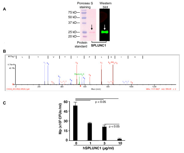
Background: Respiratory infections including Mycoplasma pneumoniae (Mp) contribute to various chronic lung diseases. We have shown that mouse short palate, lung, and nasal epithelium clone 1 (SPLUNC1) protein was able to inhibit Mp growth. Further, airway epithelial cells increased SPLUNC1 expression upon Mp infection. However, the mechanisms underlying SPLUNC1 regulation remain unknown. In the current study, we investigated if SPLUNC1 production following Mp infection is regulated through Toll-like receptor 2 (TLR2) signaling.
Methods: Airway epithelial cell cultures were utilized to reveal the contribution of TLR2 signaling including NF-κB to SPLUNC1 production upon bacterial infection and TLR2 agonist stimulation.
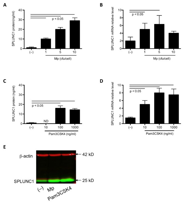
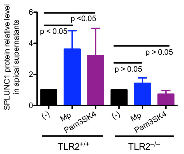
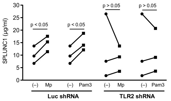
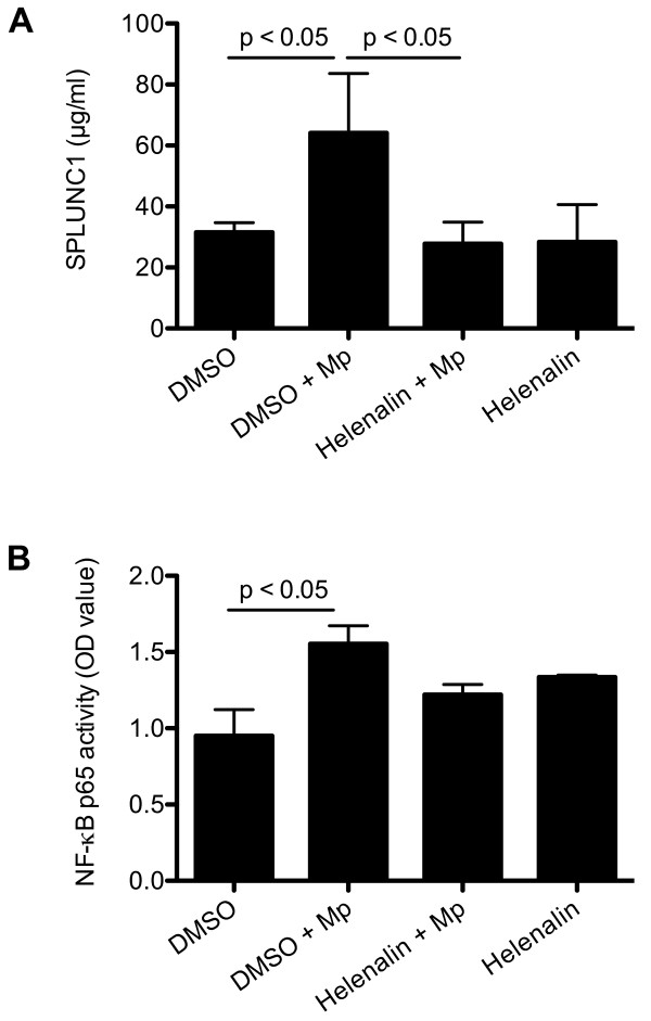
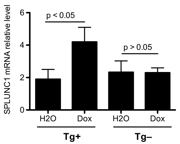
Results: Mp and TLR2 agonist Pam3CSK4 increased SPLUNC1 expression in tracheal epithelial cells from wild type, but not TLR2(-/-) BALB/c mice. RNA interference (short-hairpin RNA) of TLR2 in normal human bronchial epithelial cells under air-liquid interface cultures significantly reduced SPLUNC1 levels in Mp-infected or Pam3CSK4-treated cells. Inhibition and activation of NF-κB pathway decreased and increased SPLUNC1 production in airway epithelial cells, respectively.
Conclusions: Our data for the first time suggest that airway epithelial TLR2 signaling is pivotal in mycoplasma-induced SPLUNC1 production, thus improving our understanding of the aberrant SPLUNC1 expression in airways of patients suffering from chronic lung diseases with bacterial infections.
Figures
Similar articles
-
Function and regulation of SPLUNC1 protein in Mycoplasma infection and allergic inflammation.J Immunol. 2007 Sep 15;179(6):3995-4002. doi: 10.4049/jimmunol.179.6.3995. J Immunol. 2007. PMID: 17785838
-
Airway epithelial NF-κB activation promotes Mycoplasma pneumoniae clearance in mice.PLoS One. 2012;7(12):e52969. doi: 10.1371/journal.pone.0052969. Epub 2012 Dec 28. PLoS One. 2012. PMID: 23285237 Free PMC article.
-
MAPK/AP-1 activation mediates TLR2 agonist-induced SPLUNC1 expression in human lung epithelial cells.Mol Immunol. 2011 Dec;49(3):415-22. doi: 10.1016/j.molimm.2011.08.005. Epub 2011 Sep 6. Mol Immunol. 2011. PMID: 21899893 Free PMC article.
-
The Multifunctional Roles of Short Palate, Lung, and Nasal Epithelium Clone 1 in Regulating Airway Surface Liquid and Participating in Airway Host Defense.J Interferon Cytokine Res. 2021 Apr;41(4):139-148. doi: 10.1089/jir.2020.0141. J Interferon Cytokine Res. 2021. PMID: 33885339 Review.
-
Mammalian short palate lung and nasal epithelial clone 1 (SPLUNC1) in pH-dependent airway hydration.Int J Biochem Cell Biol. 2014 Jul;52:130-5. doi: 10.1016/j.biocel.2014.03.002. Epub 2014 Mar 13. Int J Biochem Cell Biol. 2014. PMID: 24631954 Free PMC article. Review.
Cited by
-
Whey Derivatives and Galactooligosaccharides Stimulate the Wound Healing and the Function of Human Keratinocytes through the NF-kB and FOXO-1 Signaling Pathways.Nutrients. 2022 Jul 14;14(14):2888. doi: 10.3390/nu14142888. Nutrients. 2022. PMID: 35889845 Free PMC article.
-
Hands-on workshops as an effective means of learning advanced technologies including genomics, proteomics and bioinformatics.Genomics Proteomics Bioinformatics. 2013 Dec;11(6):368-77. doi: 10.1016/j.gpb.2013.10.002. Epub 2013 Dec 6. Genomics Proteomics Bioinformatics. 2013. PMID: 24316330 Free PMC article.
-
Electronic cigarette liquid increases inflammation and virus infection in primary human airway epithelial cells.PLoS One. 2014 Sep 22;9(9):e108342. doi: 10.1371/journal.pone.0108342. eCollection 2014. PLoS One. 2014. PMID: 25244293 Free PMC article.
-
In vitro modelling of bacterial pneumonia: a comparative analysis of widely applied complex cell culture models.FEMS Microbiol Rev. 2024 Mar 1;48(2):fuae007. doi: 10.1093/femsre/fuae007. FEMS Microbiol Rev. 2024. PMID: 38409952 Free PMC article. Review.
-
Heat shock factor 1 protects against lung mycoplasma pneumoniae infection in mice.J Innate Immun. 2012;4(1):59-68. doi: 10.1159/000333089. Epub 2011 Oct 26. J Innate Immun. 2012. PMID: 22042134 Free PMC article.
References
-
- Bingle CD, Bingle L. Characterisation of the human plunc gene, a gene product with an upper airways and nasopharyngeal restricted expression pattern. Biochim Biophys Acta. 2000;1493:363–367. - PubMed
Publication types
MeSH terms
Substances
Grants and funding
LinkOut - more resources
Full Text Sources
