. 2010 Nov 5:10:341.
doi: 10.1186/1471-2148-10-341.

Evolution of xyloglucan-related genes in green plants

Affiliations

Evolution of xyloglucan-related genes in green plants

Luiz Eduardo V Del Bem et al. BMC Evol Biol. .

Abstract

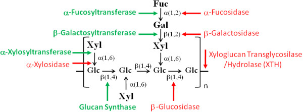
Background: The cell shape and morphology of plant tissues are intimately related to structural modifications in the primary cell wall that are associated with key processes in the regulation of cell growth and differentiation. The primary cell wall is composed mainly of cellulose immersed in a matrix of hemicellulose, pectin, lignin and some structural proteins. Xyloglucan is a hemicellulose polysaccharide present in the cell walls of all land plants (Embryophyta) and is the main hemicellulose in non-graminaceous angiosperms.

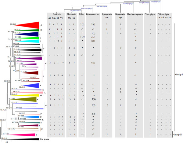
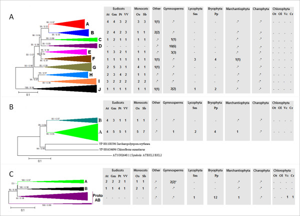
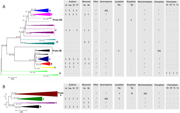
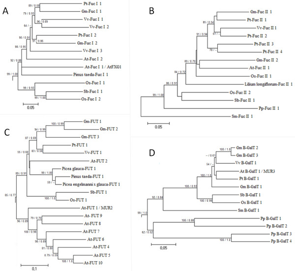
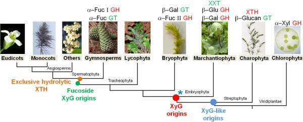
Results: In this work, we used a comparative genomic approach to obtain new insights into the evolution of the xyloglucan-related enzymatic machinery in green plants. Detailed phylogenetic analyses were done for enzymes involved in xyloglucan synthesis (xyloglucan transglycosylase/hydrolase, α-xylosidase, β-galactosidase, β-glucosidase and α-fucosidase) and mobilization/degradation (β-(1→4)-glucan synthase, α-fucosyltransferases, β-galactosyltransferases and α-xylosyl transferase) based on 12 fully sequenced genomes and expressed sequence tags from 29 species of green plants. Evidence from Chlorophyta and Streptophyta green algae indicated that part of the Embryophyta xyloglucan-related machinery evolved in an aquatic environment, before land colonization. Streptophyte algae have at least three enzymes of the xyloglucan machinery: xyloglucan transglycosylase/hydrolase, β-(1→4)-glucan synthase from the cellulose synthase-like C family and α-xylosidase that is also present in chlorophytes. Interestingly, gymnosperm sequences orthologs to xyloglucan transglycosylase/hydrolases with exclusively hydrolytic activity were also detected, suggesting that such activity must have emerged within the last common ancestor of spermatophytes. There was a positive correlation between the numbers of founder genes within each gene family and the complexity of the plant cell wall.

Conclusions: Our data support the idea that a primordial xyloglucan-like polymer emerged in streptophyte algae as a pre-adaptation that allowed plants to subsequently colonize terrestrial habitats. Our results also provide additional evidence that charophycean algae and land plants are sister groups.


Figures

Figure 1
Figure 1
Schematic representation of Xyloglucan (XyG) structure and related enzymatic activities. An oligosaccharide of XyG (XXFG) is represented. β-(1-4)-glucan synthase produce the glucan backbone, α-Xylosyl Tranferase (XXT) acts on transfer xylose residues to the main backbone, β-Galactosyl Tranferase transfers galactose residue to xylose and α-Fucosyl Transferase transfer fucose residue to galactose. Xyloglucan Transglycosylase/Hydrolase (XTH) acts on hydrolysis of XyG oligosaccharides and/or XyG transglycosylation. α-Xylosidase removes the xylose residues, β-Galactosidase removes the galactose, α-fucosidase removes the fucose and β-Glucosidase mobilizes glucose monosaccharide from the main glucan backbone.
Figure 2
Figure 2
Evolutionary profile of XTH in green plants. Phylogenetic tree showing XTH PoGOs in green plants. The topology was obtained by Neighbor-Joining method with genetic distances calculated by p-distance. The bootstrap values higher than 50% are shown for 1000 replicates along with Maximum-likelihood (ML) aLRT values higher than 0.50 (in the figure [NJ/ML]). Each triangle represents a PoGO and the amount of sequences belonging to each PoGO is proportional to triangles' size. The number of genes by species is given (At - Arabidopsis thaliana, Gm - Glycine max, Pt - Populus trichocarpa, Vv - Vitis vinifera, Os - Oryza sativa, Sb - Sorghum bicolor, Sm - Selaginella moellendorffii, Pp - Physcomitrella patens patens, Ot - Ostreococcus tauri, Ol - Ostreococcus lucimarinus, Vc - Volvox carteri, Cr - Chalmydomonas reinhardtii). Outgroup is formed by fungus sequences. Accession numbers for all genes in the tree are described in Additional File 3 and the detailed tree is shown in Additional File 2. * - No genes found within a PoGO that lacks genomic information, only ESTs. ( ) - Given number of different species within a PoGO.
Figure 3
Figure 3
Evolutionary profiles of β-galactosidase (A), XyG-active β-glucosidase (B) and α-xylosidase (C) genes in green plants. The topologies were obtained by Neighbor-Joining method with genetic distances calculated by p-distance. The bootstrap values higher than 50% are shown for 1000 replicates along with Maximum-likelihood (ML) aLRT values higher than 0.50 (in the figure [NJ/ML]). Accession numbers for all genes in the trees are described in Additional File 5 for β-galactosidase, Additional File 7 for β-glucosidase and Additional File 9 for α-xylosidase. The detailed trees are shown in Additional File 4 for β-galactosidase, Additional File 6 for β-glucosidase and Additional File 8 for α-xylosidase. The tree containing the 100 first blastp hits of AtXYL1 is shown in Additional File 10.
Figure 4
Figure 4
Evolutionary profiles of β-glucan synthase (A) and XXT (B) genes in green plants. A: CSL C (β-glucan synthase) and CSL A (β-mannan and β-glucomannan synthases) PoGOs. The topologies were obtained by Neighbor-Joining method with genetic distances calculated by p-distance. The bootstrap values higher than 50% are shown for 1000 replicates along with Maximum-likelihood (ML) aLRT values higher than 0.50 (in the figure [NJ/ML]). Accession numbers for all genes in the trees are described in Additional File 12 for β-Glucan Synthase and Additional File 14 for XXT. The detailed trees are shown in Additional File 11 for β-Glucan Synthase and Additional File 13 for XXT.
Figure 5
Figure 5
Evolutionary profiles of α-fucosidase type I (A), α-fucosidase type II (B), α-fucosyltransferase (C) and β-galactosyltransferase (D) genes in green plants. A: Arabidopsis AtXYL1 orthologs. B: Lilium longiflorum EBM II orthologs. C: Arabidopsis Mur2 orthologs. D: Arabidopsis Mur3 orthologs. The topology was obtained by Neighbor-Joining method with genetic distances calculated by p-distance. The bootstrap values higher than 50% are shown for 1000 replicates along with Maximum-likelihood (ML) aLRT values higher than 0.50 (in the figure [NJ/ML]). Accession numbers for all genes in the trees are described in the following Supplemental Tables: A and B - Additional File 16, C - Additional File 18 and 18D - Additional File 20. The detailed trees are shown in the following Additional Files: A - Additional File 15, C - Additional File 17 and 17D - Additional File 19.
Figure 6
Figure 6
Evolutionary model of XyG-related genes emergence in Viridiplantae kingdom. The model shows the ancient origins we could trace back of each XyG-related gene families and the major events of XyG evolution in plants. XyG-like (containing only glucose and xylose) emerged in streptophytes algae, XyG (containing glucose, xylose, and galactose) emerged in early embryophytes and the fucosylated XyG emerged in the last common ancestor of spermatophytes. (*) indicates the possible origins of the ancestral genes that gave rise to Spermatophytes α-Fucosyl Transferase (Mur2 orthologs) and α-Fucosylase type I (AtXYL1 orthologs). GH - Glycosyl Hydrolases and GT - Glycosyl Transferases.

Similar articles

Cited by

References

    1. Fry SC. Cross-linking of matrix polymers in the growing cell walls of angiosperms. Annual Review of Plant Physiology, Palo Alto. 1986;37:165–186. doi: 10.1146/annurev.pp.37.060186.001121. - DOI
    1. Carpita N, McCann M. In: Biochemistry and Molecular Biology of Plants. Buchanan BB, Gruissem W, Jones RL, editor. American Society of Plant Physiologists, Rockville, Maryland; 2000. The cell wall; pp. 52–108.
    1. Albert M, Werner M, Proksch P, Fry SC, Kaldenhoff R. The cell wall-modifying xyloglucan endotransglycosylase/hydrolase LeXTH1 is expressed during the defence reaction of tomato against the plant parasite Cuscuta reflexa. Plant Biol (Stuttg) 2004;6(4):402–407. doi: 10.1055/s-2004-817959. - DOI - PubMed
    1. Carpita NC, Gibeaut DM. Structural models of primary cell walls in flowering plants: consistency of molecular structure with the physical properties of the walls during growth. Plant Journal. 1993;3(1):1–30. doi: 10.1111/j.1365-313X.1993.tb00007.x. - DOI - PubMed
    1. Cosgrove DJ. Relaxation in a high-stress environment: the molecular bases of extensible cell walls and cell enlargement. Plant Cell. 1997;9(7):1031–1041. doi: 10.1105/tpc.9.7.1031. - DOI - PMC - PubMed

Publication types

LinkOut - more resources