. 2010 Nov 5:9:73.
doi: 10.1186/1475-2840-9-73.

Palmitate and insulin synergistically induce IL-6 expression in human monocytes

Affiliations

Palmitate and insulin synergistically induce IL-6 expression in human monocytes

Robert C Bunn et al. Cardiovasc Diabetol. .

Abstract

Background: Insulin resistance is associated with a proinflammatory state that promotes the development of complications such as type 2 diabetes mellitus (T2DM) and atherosclerosis. The metabolic stimuli that initiate and propagate proinflammatory cytokine production and the cellular origin of proinflammatory cytokines in insulin resistance have not been fully elucidated. Circulating proinflammatory monocytes show signs of enhanced inflammation in obese, insulin resistant subjects and are thus a potential source of proinflammatory cytokine production. The specific, circulating metabolic factors that might stimulate monocyte inflammation in insulin resistant subjects are poorly characterized. We have examined whether saturated nonesterified fatty acids (NEFA) and insulin, which increase in concentration with developing insulin resistance, can trigger the production of interleukin (IL)-6 and tumor necrosis factor (TNF)-α in human monocytes.

Methods: Messenger RNA and protein levels of the proinflammatory cytokines IL-6 and TNF-α were measured by quantitative real-time PCR (qRT-PCR) and Luminex bioassays. Student's t-test was used with a significance level of p < 0.05 to determine significance between treatment groups.

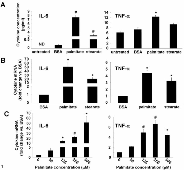
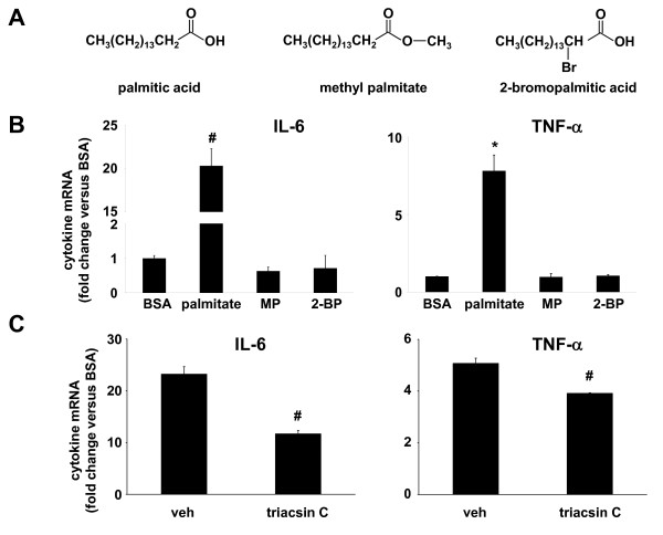
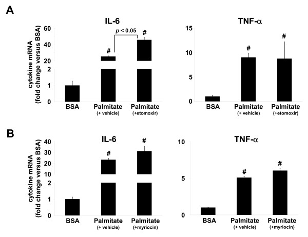
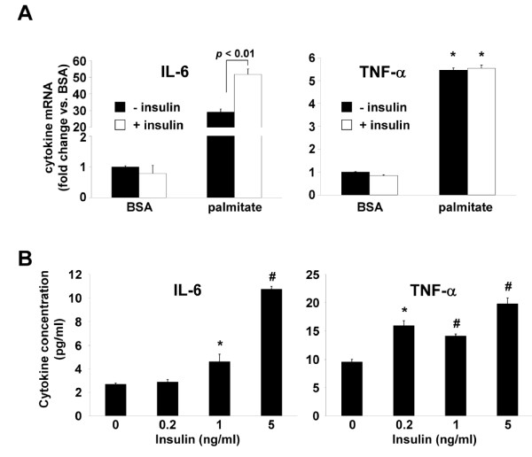
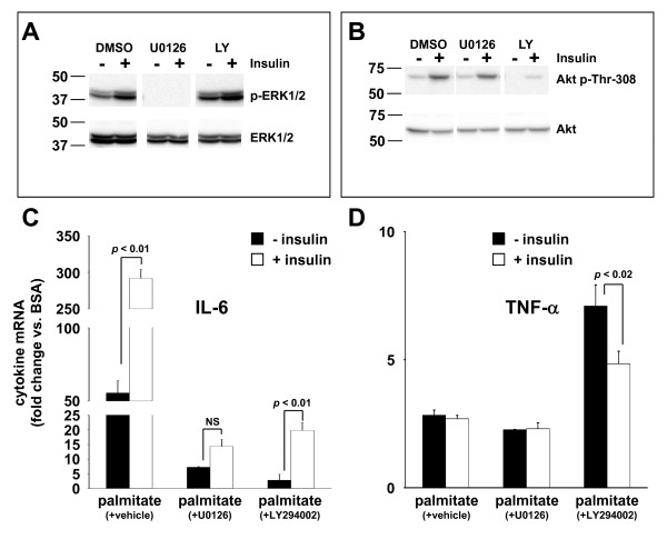
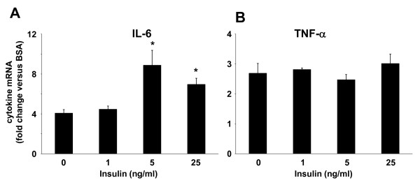
Results: Esterification of palmitate with coenzyme A (CoA) was necessary, while β-oxidation and ceramide biosynthesis were not required, for the induction of IL-6 and TNF-α in THP-1 monocytes. Monocytes incubated with insulin and palmitate together produced more IL-6 mRNA and protein, and more TNF-α protein, compared to monocytes incubated with palmitate alone. Incubation of monocytes with insulin alone did not affect the production of IL-6 or TNF-α. Both PI3K-Akt and MEK/ERK signalling pathways are important for cytokine induction by palmitate. MEK/ERK signalling is necessary for synergistic induction of IL-6 by palmitate and insulin.

Conclusions: High levels of saturated NEFA, such as palmitate, when combined with hyperinsulinemia, may activate human monocytes to produce proinflammatory cytokines and support the development and propagation of the subacute, chronic inflammatory state that is characteristic of insulin resistance. Results with inhibitors of β-oxidation and ceramide biosynthesis pathways suggest that increased fatty acid flux through the glycerolipid biosynthesis pathway may be involved in promoting proinflammatory cytokine production in monocytes.


Figures

Figure 1
Figure 1
Saturated NEFA stimulate IL-6 and TNF-α production. A, IL-6 and TNF-α concentration in 24-hour conditioned media from THP-1 cells that were untreated or stimulated with 500 μM palmitate or stearate, or NEFA- and endotoxin-free BSA. B, IL-6 and TNF-α mRNA expression in THP-1 cells stimulated as in A for 12 hours. C, IL-6 and TNF-α mRNA expression in THP-1 cells stimulated with the indicated concentrations of palmitate for 12 hours. Values are means ± SE (n = 3-4). *, p < 0.05, # p < 0.01 versus BSA treatment, ND - below the limit of detection.
Figure 2
Figure 2
Palmitate metabolism is required for the induction of IL-6 and TNF-α. A, Chemical structure of palmitic acid and non-metabolizable palmitic acid analogs methylpalmitate (MP) and 2-bromopalmitate (2-BP). B, IL-6 and TNF-α mRNA expression in THP-1 cells stimulated for 12 hours with palmitate (250 μM) or equivalent concentrations of MP or 2-BP. C, IL-6 and TNF-α mRNA expression in THP-1 cells stimulated with palmitate in the presence of vehicle (DMSO) or an inhibitor of long chain fatty acyl CoA synthetase (triacsin C, 1 μM). Values are means ± SE (n = 3). *, p < 0.05, #, p < 0.01 versus BSA.
Figure 3
Figure 3
Mitochondrial oxidation or ceramide generation is not necessary for induction of IL-6 and TNF-α by palmitate. IL-6 and TNF-α mRNA expression in THP-1 cells treated with A, etomoxir (5 μM), an inhibitor of mitochondrial fatty-acyl CoA uptake, or B, myriocin, an inhibitor of ceramide biosynthesis, for 1 hour followed by incubation with palmitate (250 μM) for 12 hours. Values are means ± SE (n = 3). #, p < 0.01 versus BSA.
Figure 4
Figure 4
Synergistic induction of IL-6 by palmitate and insulin. A, IL-6 and TNF-α mRNA expression in THP-1 cells stimulated for 30 minutes with 5 ng/ml insulin (+ insulin) or vehicle (- insulin) and then stimulated with palmitate (250 μM) or fatty acid-free BSA, for an additional 12 hours. B, IL-6 and TNF-α concentration in media from THP-1 cells stimulated for 30 minutes with the indicated concentrations of insulin and then stimulated with palmitate (125 μM) for 24 hours. Values are means ± SE (n = 3). *, p < 0.05, #, p < 0.01 versus (- insulin).
Figure 5
Figure 5
MEK/ERK and PI3K/Akt signaling pathways regulate IL-6 production. Total and phosphorylated ERK1/2 (A) or total and phosphorylated Akt (B) were analyzed by Western blotting of cellular extracts from control (-) and insulin-stimulated (+) THP-1 cells that were pre-treated with vehicle (DMSO), MEK1/2 inhibitor (U0126), or PI3K inhibitor (LY294002). Western blots representative of three independent experiments are shown. IL-6 (C) and TNF-α (D) mRNA expression determined by qRT-PCR in THP-1 cells treated for 1 hour as in (A) followed by stimulation for 30 minutes with 5 ng/ml insulin (+ insulin) or vehicle (- insulin), in the presence of vehicle (DMSO), and then stimulated with palmitate (250 μM) or fatty acid-free BSA, for an additional 24 hours. Values are means ± SE (n = 3) expressed relative to the expression level of these cytokines in BSA-treated cells.
Figure 6
Figure 6
Synergistic induction of IL-6 mRNA in primary human monocytes. Monocytes isolated by adherence from human PBMC fraction of whole blood were incubated for 30 minutes with varying concentrations of insulin, followed by 24 hour incubation with 500 μM palmitate or BSA. IL-6 (A) and TNF-α (B) mRNAs were determined by qRT-PCR. Values are means ± SE (n = 3) expressed relative to the expression level of these cytokines in BSA-treated cells. *, p < 0.05 versus (- insulin).

Similar articles

Cited by

References

    1. Reaven GM. Pathophysiology of insulin resistance in human disease. Physiol Rev. 1995;75:473–486. - PubMed
    1. Razani B, Chakravarthy MV, Semenkovich CF. Insulin resistance and atherosclerosis. Endocrinol Metab Clin North Am. 2008;37:603–621. doi: 10.1016/j.ecl.2008.05.001. viii. - DOI - PMC - PubMed
    1. Shoelson SE, Herrero L, Naaz A. Obesity, inflammation, and insulin resistance. Gastroenterology. 2007;132:2169–2180. doi: 10.1053/j.gastro.2007.03.059. - DOI - PubMed
    1. Yudkin JS, Kumari M, Humphries SE, Mohamed-Ali V. Inflammation, obesity, stress and coronary heart disease: is interleukin-6 the link? Atherosclerosis. 2000;148:209–214. doi: 10.1016/S0021-9150(99)00463-3. - DOI - PubMed
    1. Hotamisligil GS, Shargill NS, Spiegelman BM. Adipose expression of tumor necrosis factor-alpha: direct role in obesity-linked insulin resistance. Science. 1993;259:87–91. doi: 10.1126/science.7678183. - DOI - PubMed

Publication types

MeSH terms