. 2010 Nov 12;87(5):701-7.
doi: 10.1016/j.ajhg.2010.10.006. Epub 2010 Nov 4.

Mutations in myosin light chain kinase cause familial aortic dissections

Affiliations

Mutations in myosin light chain kinase cause familial aortic dissections

Li Wang et al. Am J Hum Genet. .

Erratum in

  • Am J Hum Genet. 2011 Apr 8;88(4):516

Abstract

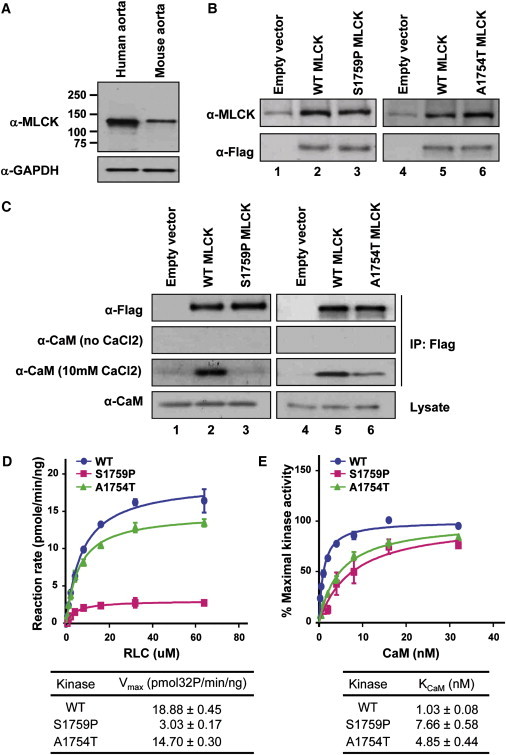
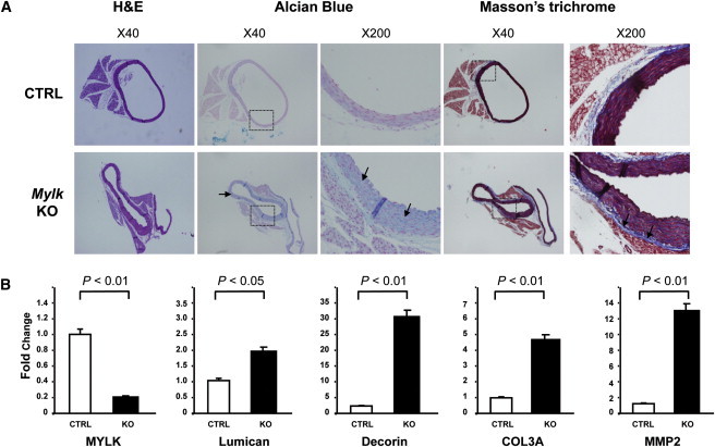
Mutations in smooth muscle cell (SMC)-specific isoforms of α-actin and β-myosin heavy chain, two major components of the SMC contractile unit, cause familial thoracic aortic aneurysms leading to acute aortic dissections (FTAAD). To investigate whether mutations in the kinase that controls SMC contractile function (myosin light chain kinase [MYLK]) cause FTAAD, we sequenced MYLK by using DNA from 193 affected probands from unrelated FTAAD families. One nonsense and four missense variants were identified in MYLK and were not present in matched controls. Two variants, p.R1480X (c.4438C>T) and p.S1759P (c.5275T>C), segregated with aortic dissections in two families with a maximum LOD score of 2.1, providing evidence of linkage of these rare variants to the disease (p = 0.0009). Both families demonstrated a similar phenotype characterized by presentation with an acute aortic dissection with little to no enlargement of the aorta. The p.R1480X mutation leads to a truncated protein lacking the kinase and calmodulin binding domains, and p.S1759P alters amino acids in the α-helix of the calmodulin binding sequence, which disrupts kinase binding to calmodulin and reduces kinase activity in vitro. Furthermore, mice with SMC-specific knockdown of Mylk demonstrate altered gene expression and pathology consistent with medial degeneration of the aorta. Thus, genetic and functional studies support the conclusion that heterozygous loss-of-function mutations in MYLK are associated with aortic dissections.


Figures

Figure 1
Figure 1
Identification of MYLK as a Causative Gene Leading to Familial TAAD (A) This panel shows the two families with MYLK mutations, p.S1759P and p.R1480X, which segregate with TAAD. The disease status and mutation status of individuals are indicated in the figure key. Current age or age at death for affected individuals and the current age for unaffected individuals are indicated under the individual ID. (B) MYLK encodes three products from independent promoters: long form, short form, and telokin. The alterations in black indicate polymorphisms that are present in controls. The alterations in blue are absent in ethnically matched controls, but segregation with disease could not be tested. These changes do not affect conserved amino acids and do not disrupt the kinase domain. The alterations shown in red are predicted to affect kinase activity. (C) Three-dimensional structures of the MLCK kinase domain and the CaM-binding sequence are presented in green and yellow, respectively. The remainder of the structure is shown in gray. The CaM-binding sequence forms a basic α-helix secondary structure containing Alaline-1754 and Serine-1759, shown in red.
Figure 2
Figure 2
Expression of MLCK in Aorta and Assessment of Kinase Activity in MYLK Mutants (A) Long-form and short-form MLCK were examined in human and mouse aortas. Only a 130 kDa protein band was detected in extracts from aortic tissues. Positions of molecular-weight markers are indicated at the side of the panel. (B) COS7 cells were transfected with an empty vector (lane 1) or with vectors expressing Flag-tagged, wild-type short-form MLCK (lane 2), or S1759P short-form MLCK (lane 3). After 48 hr, whole cell lysates were collected for detection of target proteins by immunoblotting with anti-MLCK and anti-Flag. In contrast to lane 1, Flag-tagged short-form MLCK proteins were detected in lanes 2 and 3. Additional COS-7 cells were transfected with empty vectors (lane 4) or with vectors expressing Flag-tagged, wild-type short-form MLCK (lane 5) or A1754T short-form MLCK (lane 6). The target proteins were also detected with anti-MLCK and anti-Flag. (C) Binding of CaM to MLCK mutants. COS7 cell lysates expressing empty vectors (lane 1), wild-type short-form MLCK (lane 2), and S1759P short-form MLCK (lane 3) were pulled down with the use of anti-Flag. Immunoprecipitates were analyzed by immunoblotting with the use of anti-CaM. In the presence of 10 mM CaCl2, CaM was coimmunoprecipitated with wild-type short-form MLCK but not with S1759P short-form MLCK. In the absence of CaCl2, no α-CaM signal was detected. Additional COS7 cell lysates expressing empty vectors (lane 4), wild-type short-form MLCK (lane 5), and A1754T short-form MLCK (lane 6) were pulled down with the use of anti-Flag and blotted with anti-CaM. The CaM band in lane 5 is more strongly detected than the band in lane 6 in the presence of 10 mM CaCl2. (D) Assessment of kinase activity of wild-type and mutant MLCK proteins. The rate of 32P incorporation into RLC was measured. The maximal activities of WT MLCK (blue circle), A1754T MLCK (green triangle), and S1759P MLCK (red square) were obtained at different RLC concentrations. The data points represented the mean ± standard error of three or more determinations. The data were fit to the Michaelis-Menten equation for calculation of the Vmax values. (E) CaM activation of wild-type and mutant MLCK proteins. The relative percentage of maximal kinase activity of WT MLCK (blue circle), A1754T MLCK (green triangle), and S1759P MLCK (red square) was plotted versus various CaM concentrations. The data were presented as mean ± standard error from three experiments. The data were fit to the Michaelis-Menten equation to obtain the KCaM value.
Figure 3
Figure 3
Aortic Pathology from Two Patients with the MLCK mutation p.S1759P, TAA026:II:2 and TAA026:II:3, and an Unaffected Control (A) Movat staining of aortas from patients shows medial degeneration of the aorta, as shown by increased proteoglycan deposition (blue, arrows) and elastic-fiber fragmentation and loss (black). Von Willebrand factor (marker of endothelial cells) immunostaining of the aorta (red) was performed to demonstrate the increased vascularity in the medial layer in TAA026:II2 and TAA026:II3 (arrows) as compared to the control aorta. (B) Quantification of the number of arteries in the aortic media from patients with MYLK mutations and controls. The average number of arteries per field from the patients' aortic media is significantly higher than that in the control aortas. Data are expressed as mean ± standard error of the mean, and p values are indicated.
Figure 4
Figure 4
Pathology of the Ascending Aorta with Smooth-Muscle-Specific Deficiency of MLCK in Mice Mylkf/f mice were bred to homozygosity and crossed with a transgenic mouse line expressing tamoxifen-activated Cre under the control of the smooth-muscle myosin heavy chain promoter.MlckSMKO male mice 12–13 weeks old and littermate controls harboring the Cre transgene in mixed 129/B6 backgrounds were used for this study. (A) Paraffin-embedded ascending aortic tissues from control (n = 3) and SM-Mylk-KO (n = 4) mice were sectioned and stained for proteoglycans and collagen. The upper panels are from the control mice (CTRL), and the lower panels are from the tamoxifen-treated Sm-Mylk-KO mice (KO). The specific stains and magnifications are indicted above the panels. Alcian blue staining indicates increased proteoglycan accumulation (blue) in the SM-Mylk-KO mice compared with controls (arrows). Masson's trichrome staining indicates increased collagen fibers (blue) in the adventitia (arrows) in the SM-MYLK-KO mice compared with controls. (B) Quantitative PCR was used to determine changes in expression levels of MYLK, MMP2, lumican, decorin, and type III collagen. Expression analysis was performed with the use of RNA extracted from ascending aortas of control (n = 4) and Sm-Mylk-KO (n = 4) mice. The analysis shows decreased MYLK expression, whereas MMP2 messages increased 13-fold, lumican messages increased 2-fold, decorin messages increased 32-fold, and the type III procollagen (COL3A1) messages increased 5-fold. Gene-expression levels are standardized to GAPDH messages. The relative expression values were determined via the ΔΔCt method, and assays were performed in triplicate. Data are expressed as mean ± standard deviation.

Comment in

Similar articles

Cited by

References

    1. Kamm K.E., Stull J.T. Dedicated myosin light chain kinases with diverse cellular functions. J. Biol. Chem. 2001;276:4527–4530. - PubMed
    1. He W.Q., Peng Y.J., Zhang W.C., Lv N., Tang J., Chen C., Zhang C.H., Gao S., Chen H.Q., Zhi G. Myosin light chain kinase is central to smooth muscle contraction and required for gastrointestinal motility in mice. Gastroenterology. 2008;135:610–620. - PMC - PubMed
    1. Schubert R., Lidington D., Bolz S.S. The emerging role of Ca2+ sensitivity regulation in promoting myogenic vasoconstriction. Cardiovasc. Res. 2008;77:8–18. - PubMed
    1. Welsh D.G., Nelson M.T., Eckman D.M., Brayden J.E. Swelling-activated cation channels mediate depolarization of rat cerebrovascular smooth muscle by hyposmolarity and intravascular pressure. J. Physiol. 2000;527:139–148. - PMC - PubMed
    1. Isotani E., Zhi G., Lau K.S., Huang J., Mizuno Y., Persechini A., Geguchadze R., Kamm K.E., Stull J.T. Real-time evaluation of myosin light chain kinase activation in smooth muscle tissues from a transgenic calmodulin-biosensor mouse. Proc. Natl. Acad. Sci. USA. 2004;101:6279–6284. - PMC - PubMed

Publication types

Substances