. 2011 Feb;32(5):1317-26.
doi: 10.1016/j.biomaterials.2010.10.006. Epub 2010 Nov 4.

Human urine-derived stem cells seeded in a modified 3D porous small intestinal submucosa scaffold for urethral tissue engineering

Affiliations

Human urine-derived stem cells seeded in a modified 3D porous small intestinal submucosa scaffold for urethral tissue engineering

Shaofeng Wu et al. Biomaterials. 2011 Feb.

Abstract

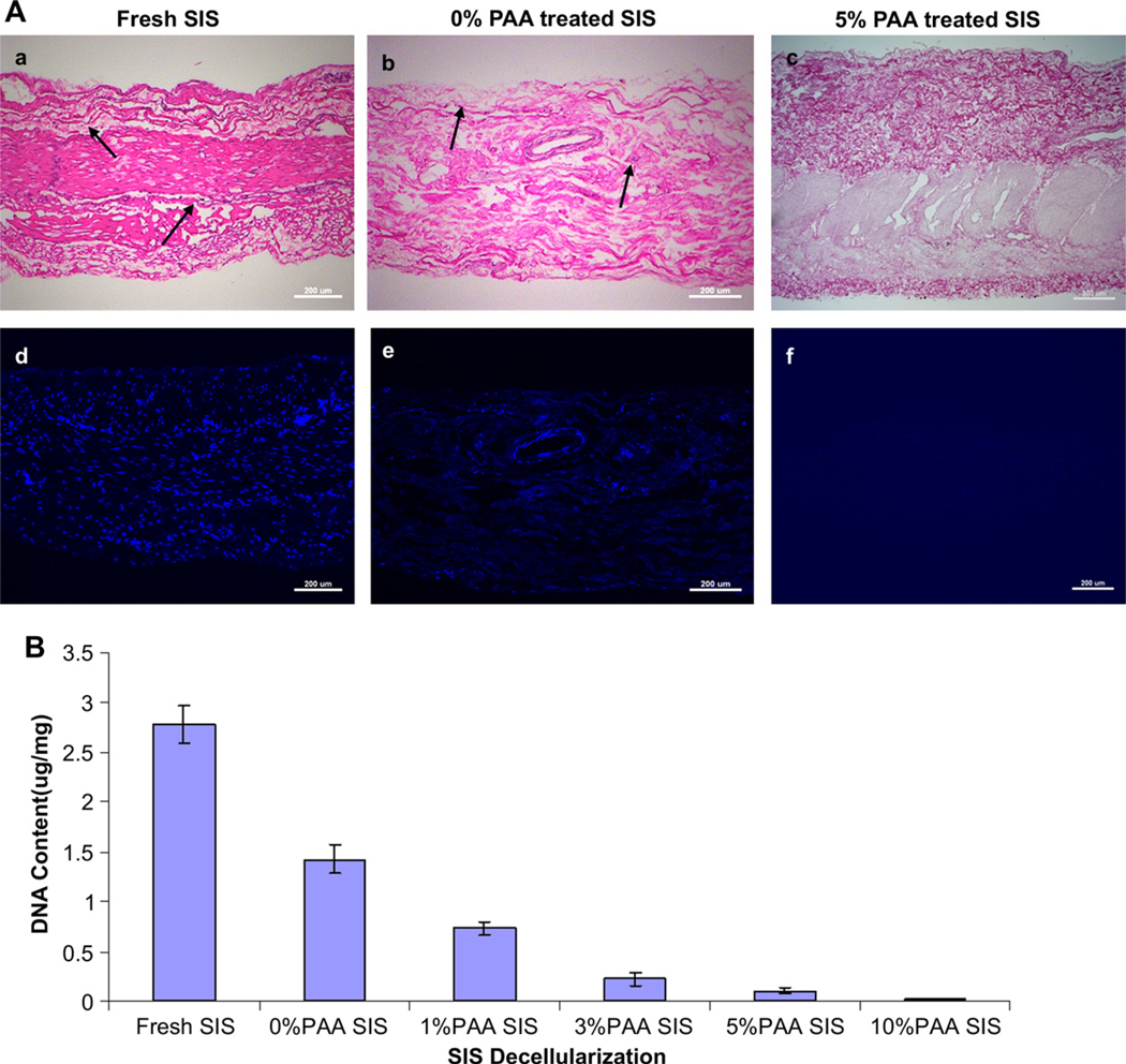
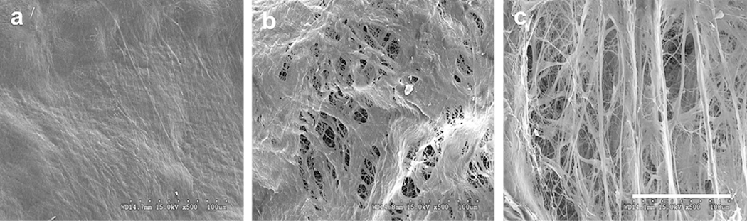
The goal of this study was to determine whether urothelial cells (UC) and smooth muscle cells (SMC) derived from the differentiation of urine-derived stem cells (USC) could be used to form engineered urethral tissue when seeded on a modified 3-D porous small intestinal submucosa (SIS) scaffold. Cells were obtained from 12 voided urine samples from 4 healthy individuals. USC were isolated, characterized and induced to differentiate into UC and SMC. Fresh SIS derived from pigs was decellularized with 5% peracetic acid (PAA). Differentiated UC and SMC derived from USC were seeded onto SIS scaffolds with highly porous microstructure in a layered co-culture fashion and cultured under dynamic conditions for one week. The seeded cells formed multiple uniform layers on the SIS and penetrated deeper into the porous matrix during dynamic culture. USC that were induced to differentiate also expressed UC markers (Uroplakin-III and AE1/AE3) or SMC markers (α-SM actin, desmin, and myosin) after implantation into athymic mice for one month, and the resulting tissues were similar to those formed when UC and SMC derived from native ureter were used. In conclusion, UC and SMC derived from USC could be maintained on 3-D porous SIS scaffold. The dynamic culture system promoted 3-D cell-matrix ingrowth and development of a multilayer mucosal structure similar to that of native urinary tract tissue. USC may serve as an alternative cell source in cell-based tissue engineering for urethral reconstruction or other urological tissue repair.


Figures

Fig. 1.
Fig. 1.
Comparison of gross morphology of decellularized SIS with PAA treatment. (A) H&E (a,b,c) and DAPI staining (d,e,f) on cross sections of fresh SIS (a,d), 0% PAA (i.e. distill water with non-PAA treated) treated SIS (b,e) and 5% PAA-treated SIS (c,f). Cells and cell nuclei are visible in fresh SIS tissue (arrows; a,d) whereas a rapid decrease in cellular contents was visible with 0% PAA treatment (decellularization alone) (arrows; b,e). However, complete removal of all cellular components was achieved using 5% PAA treatment (c,f). An increase in porosity of the micro-architecture of SIS was visible after PAA treatment (c,f). Scale bar represents 200 um. (B) Effect of PAA on DNA clearance. DNA was extracted from fresh SIS and decellularized SIS prior to and after PPA treatment. The DNA values were plotted after normalizing to initial wet weight of the sample. A significant clearance of DNA content was observed with increased PAA concentrations.
Fig. 2.
Fig. 2.
Electron micrograph of mucosal side of decellularized SIS with PAA. (a) Untreated (fresh) SIS, (b) Decellularized SIS (0% PAA treatment) and (c) Decellularized SIS (5% PAA). A remarkable high level of porosity was achieved after treatment with 5% PAA when compared to the 0% PAA treatment. Scale bar = 100 um.
Fig. 3.
Fig. 3.
Effect of PAA concentration on cell penetration and proliferation. SIS were treated with 0% and 5% PAA prior to seeding with ureter SMC and UC for 14 Days. Sections were stained with Masson’s Trichrome (top panel) and DAPI (bottom panel). Cell–matrix ingrowth in 5% PAA-treated SIS were significantly higher (arrows, indicating presence of cells at different depths) than that of the 0% PAA treatment. UC and SMC layers are marked. The vertical black line in the top panels on the left side indicates the scaffold thickness. Scale bar represents 100 um.
Fig. 4.
Fig. 4.
Effect of culture condition on cell penetration and proliferation. A) Masson’s Trichrome staining (top panel) and DAPI staining (bottom panel) performed on 5% PAA-treated SIS, seeded with ureter SMC and UC under static or dynamic (10 or 40 RPM) culture condition for 14 d. Cell penetration (lower arrow) and proliferation (upper arrow) was significantly improved with dynamic culture condition as compared to static culture condition. The vertical black line in the top panels denotes scaffold thickness. Inset images shows lower magnification of the entire thickness of the scaffold. Scale bar represents 100 um. B) Analysis of cell growth. Ureter SMC and UC seeded SIS were analyzed for cell growth using the MTT assay. OD values were recorded after reading the plate at 450 nm. The graph shows cells grown in the PAA-treated (5%) and the untreated (0%) under different static and dynamic (10 and 40 RPM) culture condition. Irrespective of the culture conditions, the treatment with PAA caused a significant growth of cells compared to the non-treatment (0%) (p < 0.05).
Fig. 5.
Fig. 5.
Tracking of human cell-seeded SIS in vitro and in vivo. A) Masson’s trichrome staining, as shown on the left at 200×, depicts cellular (SMC) penetration (black arrows) and proliferation (black arrowheads) as well as UC layers (white arrows). The in vivo image at 400× shows appearance of vessel-like structures (black arrows). B) Detection of human ureter cells by immunostaining with human nuclear antibody (NuMA, in green) in layered co-culture of ureter UC/SMC (as controls in the left column) and SMC-USC along with UC-USC in the right column) in vitro (top panel) as well as in vivo (bottom panel), 4 weeks after implantation. Arrows indicate representative strong positively labeled implanted human cells. As the cell nuclei were counterstained with PI, human cells appear yellow due to merging of the green and red signal. Scale bar represents 50 um.
Fig. 6.
Fig. 6.
Immunofluorescent staining of urothelial-differentiated USC in vitro and in vivo. Tissue sections of in vivo and in vitro cultured cells were immunostained (green) using A) pancytokeratin (AE1/AE3) and B) Uroplakin-III antibodies. USC were differentiated in vitro with EGF for 14 days by using dynamic culture condition (10 RPM) followed by subcutaneous implantation into nude mice for a further 4 weeks. The nuclei were counterstained with PI (red). The left panel indicates in vitro and in vivo co-cultures of UC and SMC as a positive control. Arrows show a few representative cells that are positively stained. Scale bar represents 50 um.
Fig. 7.
Fig. 7.
Immunofluorescent staining of myogenic-differentiated USC in vitro and in vivo. Tissue sections of in vitro (top panel) and in vivo (bottom panel) cultured cells that were immunostained (green or red) using A) α-SM actin, B) Desmin and C) Myosin antibodies. USC were differentiated in vitro using PDGF and TGF for 14 days under dynamic condition (10 RPM), following by subcutaneously implantation into nude mice for a further 4 weeks (right column). In vitro and in vivo seeded UC/SMC scaffolds (left column) were used as controls. Positively stained cells for α SM actin and Myosin appear green (FITC), whereas staining for Desmin appears red (Rhodamine). Nuclei were counterstained with PI (red) or DAPI (blue). Arrows show a few representative cells that are positively stained. Scale bar represents 50 um.

References

    1. Atala A, Bauer SB, Soker S, Yoo JJ, Retik AB. Tissue-engineered autologous bladders for patients needing cystoplasty. Lancet 2006;367:1241–6. - PubMed
    1. Zhang Y, McNeill E, Tian H, Soker S, Andersson KE, Yoo JJ, et al. Urine derived cells are a potential source for urological tissue reconstruction. J Urol 2008;180:2226–33. - PubMed
    1. Bharadwaj S, Wu S, Rohozinski J, Furth M, Lan X, Atala A, et al. Multipotential differentiation of human urine-derived stem cells. J Tissue Eng Regen Med 2009;6:S293.
    1. Djordjevic ML, Majstorovic M, Stanojevic D, Bizic M, Ducic S, Kojovic V, et al. One-stage repair of severe hypospadias using combined buccal mucosa graft and longitudinal dorsal skin flap. Eur J Pediatr Surg 2008;18:427–30. - PubMed
    1. Mungadi IA, Ugboko VI. Oral mucosa grafts for urethral reconstruction. Ann Afr Med 2009;8:203–9. - PubMed

Substances