

In vivo confocal microscopy of trachoma in relation to normal tarsal conjunctiva
- PMID: 21055819
- PMCID: PMC3267042
- DOI: 10.1016/j.ophtha.2010.08.029
In vivo confocal microscopy of trachoma in relation to normal tarsal conjunctiva
Abstract
Objective: To describe the in vivo confocal microscopy (IVCM) appearances of the tarsal conjunctiva in trachoma compared with the appearance of healthy conjunctiva and to develop grading systems for IVCM examination of the tarsal conjunctiva for use in future studies on trachoma and other conjunctival diseases.
Design: Prospective observational study.
Participants: In vivo confocal microscopy examination was performed on 302 clinically normal adults, 16 clinically normal children, 750 adults with trachomatous conjunctival scarring, and 25 children with active trachoma.
Methods: Clinical evaluation was performed with ×2.5 loupes, and IVCM examination of the upper tarsal conjunctiva was carried out with a Heidelberg Retina Tomograph 3 with the Rostock Cornea Module (Heidelberg Engineering GmbH, Dossenheim, Germany).
Main outcome measures: In vivo confocal microscopy images were analyzed for cellular and tissue changes associated with trachomatous inflammation and scarring compared with healthy subjects.
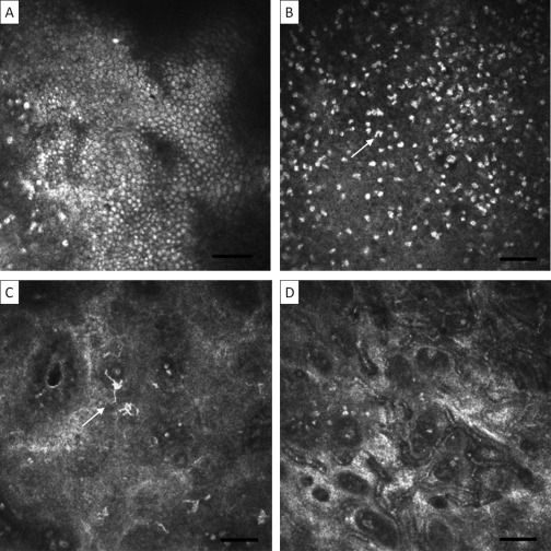
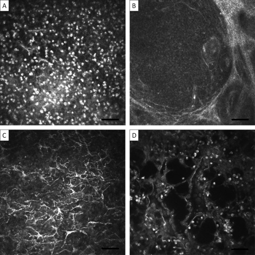
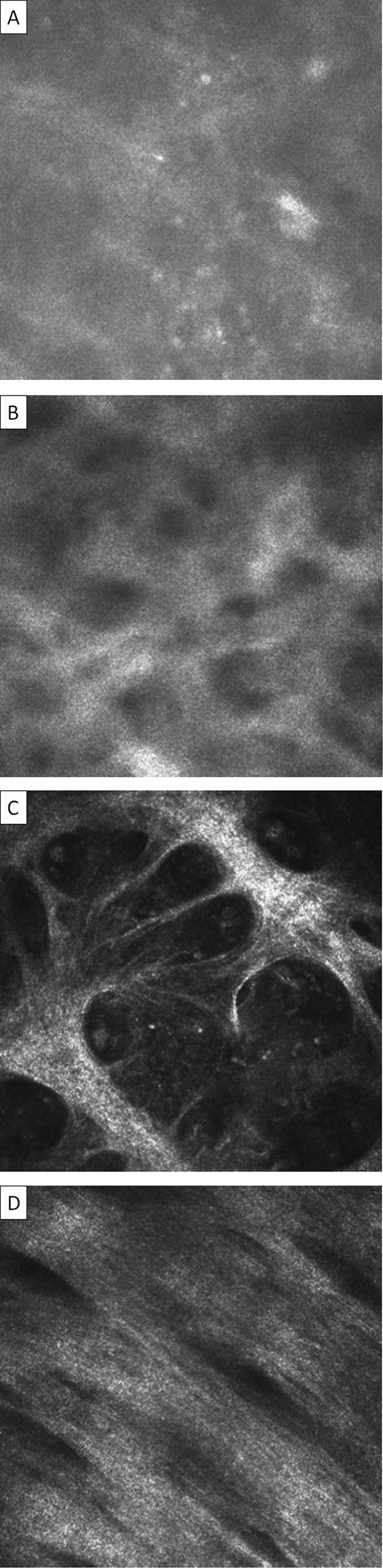
Results: Trachomatous subjects with follicular and papillary inflammation had an increased inflammatory cellular infiltrate, including dendritiform cells, discrete follicular and papillary structures, and cystic lacunae suggestive of tissue edema. Trachomatous conjunctival scarring was seen with IVCM as organization of the subepithelial connective tissue into bands/sheets. Grading systems for inflammatory changes and scarring were developed, with the system for scarring showing good interobserver agreement with an intraclass coefficient of 0.88.
Conclusions: In vivo confocal microscopy provides a powerful tool for examining the ocular surface. Numerous cellular and tissue changes were observed in subjects with trachoma, the first time IVCM has been applied to this disease. These changes both complement and add to previous histologic analyses. In vivo confocal microscopy promises to provide new insights into the pathogenesis of trachoma and other conjunctival diseases.
Copyright © 2011 American Academy of Ophthalmology. Published by Elsevier Inc. All rights reserved.
Figures
Similar articles
-
In vivo confocal microscopy and histopathology of the conjunctiva in trachomatous scarring and normal tissue: a systematic comparison.Br J Ophthalmol. 2013 Oct;97(10):1333-7. doi: 10.1136/bjophthalmol-2013-303126. Epub 2013 Aug 6. Br J Ophthalmol. 2013. PMID: 23922266 Free PMC article.
-
A review of the application of in-vivo confocal microscopy on conjunctival diseases.Eye Vis (Lond). 2024 Nov 1;11(1):43. doi: 10.1186/s40662-024-00409-x. Eye Vis (Lond). 2024. PMID: 39482793 Free PMC article. Review.
-
In vivo confocal microscopy in scarring trachoma.Ophthalmology. 2011 Nov;118(11):2138-46. doi: 10.1016/j.ophtha.2011.04.014. Epub 2011 Sep 13. Ophthalmology. 2011. PMID: 21920608 Free PMC article.
-
In vivo confocal microscopy and trachomatous conjunctival scarring: Predictors for clinical progression.Clin Exp Ophthalmol. 2020 Dec;48(9):1152-1159. doi: 10.1111/ceo.13843. Epub 2020 Sep 16. Clin Exp Ophthalmol. 2020. PMID: 32798249
-
Immunology of trachomatous conjunctivitis.Bull Soc Belge Ophtalmol. 2001;(280):73-96. Bull Soc Belge Ophtalmol. 2001. PMID: 11486468 Review.
Cited by
-
Conjunctival transcriptome in scarring trachoma.Infect Immun. 2011 Jan;79(1):499-511. doi: 10.1128/IAI.00888-10. Epub 2010 Oct 11. Infect Immun. 2011. PMID: 20937763 Free PMC article.
-
Conjunctival Microbiome-Host Responses Are Associated With Impaired Epithelial Cell Health in Both Early and Late Stages of Trachoma.Front Cell Infect Microbiol. 2019 Aug 21;9:297. doi: 10.3389/fcimb.2019.00297. eCollection 2019. Front Cell Infect Microbiol. 2019. PMID: 31552195 Free PMC article.
-
In vivo confocal microscopy and histopathology of the conjunctiva in trachomatous scarring and normal tissue: a systematic comparison.Br J Ophthalmol. 2013 Oct;97(10):1333-7. doi: 10.1136/bjophthalmol-2013-303126. Epub 2013 Aug 6. Br J Ophthalmol. 2013. PMID: 23922266 Free PMC article.
-
Trachoma: protective and pathogenic ocular immune responses to Chlamydia trachomatis.PLoS Negl Trop Dis. 2013;7(2):e2020. doi: 10.1371/journal.pntd.0002020. Epub 2013 Feb 14. PLoS Negl Trop Dis. 2013. PMID: 23457650 Free PMC article. Review.
-
A review of the application of in-vivo confocal microscopy on conjunctival diseases.Eye Vis (Lond). 2024 Nov 1;11(1):43. doi: 10.1186/s40662-024-00409-x. Eye Vis (Lond). 2024. PMID: 39482793 Free PMC article. Review.
References
-
- Mariotti S.P., Pascolini D., Rose-Nussbaumer J. Trachoma: global magnitude of a preventable cause of blindness. Br J Ophthalmol. 2009;93:563–568. - PubMed
-
- Guthoff R.F., Baudouin C., Stave J. Springer; Berlin: 2006. Atlas of Confocal Laser Scanning In-vivo Microscopy in Ophthalmology; pp. 89–90.
-
- Chiou A.G., Kaufman S.C., Kaufman H.E., Beuerman R.W. Clinical corneal confocal microscopy. Surv Ophthalmol. 2006;51:482–500. - PubMed
-
- Mastropasqua L., Nubile M., Lanzini M. Epithelial dendritic cell distribution in normal and inflamed human cornea: in vivo confocal microscopy study. Am J Ophthalmol. 2006;142:736–744. - PubMed
Publication types
MeSH terms
Grants and funding
LinkOut - more resources
Full Text Sources
