. 2011 Feb;140(2):667-75.
doi: 10.1053/j.gastro.2010.10.056. Epub 2010 Nov 4.

Protease inhibitor-resistant hepatitis C virus mutants with reduced fitness from impaired production of infectious virus

Affiliations

Protease inhibitor-resistant hepatitis C virus mutants with reduced fitness from impaired production of infectious virus

Tetsuro Shimakami et al. Gastroenterology. 2011 Feb.

Abstract

Background & aims: Several small molecule inhibitors of the hepatitis C virus (HCV) nonstructural protein (NS) 3/4A protease have advanced successfully to clinical trials. However, the selection of drug-resistant mutants is a significant issue with protease inhibitors (PIs). A variety of amino acid substitutions in the protease domain of NS3 can lead to PI resistance. Many of these significantly impair the replication fitness of HCV RNA replicons. However, it is not known whether these mutations also adversely affect infectious virus assembly and release, processes in which NS3 also participates.

Methods: We studied the impact of 25 previously identified PI-resistance mutations on the capacity of genotype 1a H77S RNA to replicate in cell culture and produce infectious virus.

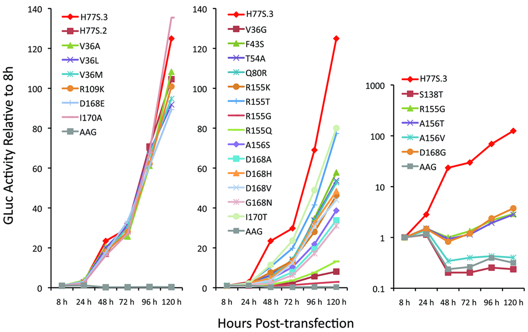
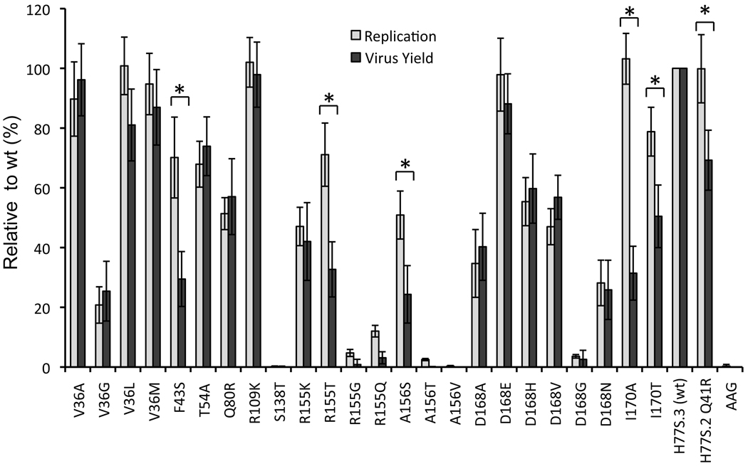
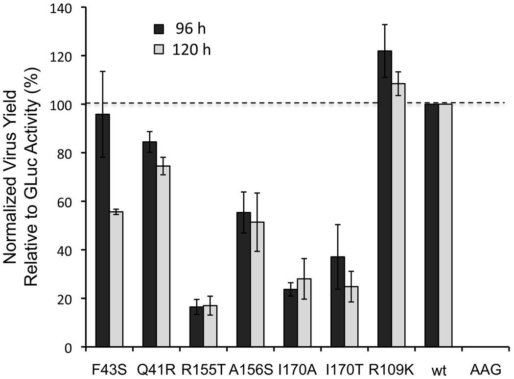
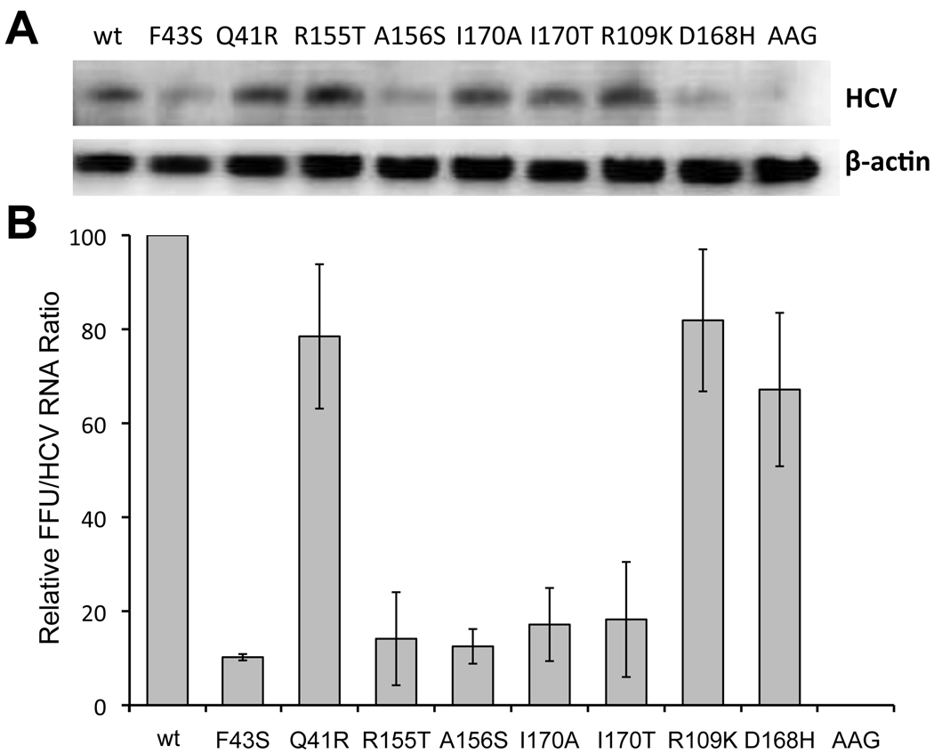
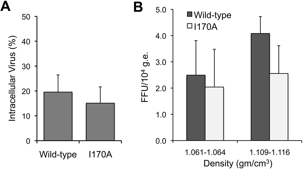
Results: Most PI-resistance mutations resulted in moderate loss of replication competence, although several (V36A/L/M, R109K, and D168E) showed fitness comparable to wild type, whereas others (S138T and A156V) were severely impaired both in RNA replication and infectious virus production. Although reductions in RNA replication capacity correlated with decreased yields of infectious virus for most mutations, a subset of mutants (Q41R, F43S, R155T, A156S, and I170A/T) showed greater impairment in their ability to produce virus than predicted from reductions in RNA replication capacity. Detailed examination of the I170A mutant showed no defect in release of virus from cells and no significant difference in specific infectivity of extracellular virus particles.

Conclusions: Replicon-based assays might underestimate the loss of fitness caused by PI-resistance mutations, because some mutations in the NS3 protease domain specifically impair late steps in the viral life cycle that involve intracellular assembly of infectious virus.


Figures

Figure 1
Figure 1. Impact of drug-resistance mutations on genotype 1a HCV RNA replication
Medium was collected and replaced at 8, 24, 48, 72, 96, 120h after transfection of H77S.3/GLuc2A RNAs carrying the indicated mutations, and GLuc activity determined at each point in time. Results were normalized to the 8h GLuc activity, and represent the mean of triplicate samples and are representative of multiple experiments. The mutants are grouped in 3 panels, with increasingly negative impact on replication capacity from left to right.
Figure 2
Figure 2. Impact of drug-resistance mutations on HCV RNA replication and infectious virus production
GLuc activity secreted by RNA-transfected cells at 96 vs. 8 h, as shown in Fig. 1, was normalized to that of the wild-type H77S.3 RNA (100% = 66 ± 19 fold-increase, lightly shaded bars) and plotted adjacent to infectious virus yield (combined data from 72 and 96 hr endpoints) from each mutant, similarly normalized to H77S.3 RNA (100% = 3.4 ± 1.3×10³ FFU/ml, solid bars). The data shown represent the mean ± S.D. from at least three (replication) or four (virus yield) independent experiments. An asterix [*] designates mutants for which the difference between the relative capacities to replicate as RNA and to produce infectious virus is significant by student’s t-test (p<0.001). “wt” refers to wild type.
Figure 3
Figure 3. Infectious virus yield versus GLuc expression from H77S.3/GLuc2A mutants
Supernatant culture fluids were assayed for GLuc activity as well as infectious virus 96h and 120h following transfection of H77S.3/GLuc2A and related mutant RNAs. Results are shown as the ratio of infectious virus to GLuc activity, normalized to wild-type H77S.3/GLuc2A RNA (100% = 90 ± 12 FFU/ml/103 light units (L.U.) at 96h, and 88 ± 11 FFU/ml/103 L.U. at 120h). Results are means ± S.D. from 3 experiments.
Figure 4
Figure 4. Infectious virus yield vs. RNA abundance of H77S.3 mutants
H77S.3 and related RNAs containing the indicated mutations were transfected into Huh-7.5 cells. (A) Cells were lysed and total RNA extracted at 96h, then subjected to northern blotting with HCV-and β-actin-specific probes. (B) Ratio of extracellular infectious virus at 96h to intracellular RNA abundance quantified by phosphor image analysis of northern blots (HCV/β-actin RNA), normalized to yields from wild-type H77S.3 RNA (1.2 ± 0.52×10³ FFU/ml). Results shown are means ± range from duplicate experiments.
Figure 5
Figure 5
A PI resistance mutation causes a defect in assembly of infectious virus. (A) Intracellular infectious virus titer as the percent of extracellular wild-type (H77S.3) and I170A virus 96h after RNA transfection. Results are means ± S.D. (B) Specific infectivity of the major species of extracellular wild-type (H77S.3) and I170A virus particles isolated by equilibrium gradient centrifugation. Results are mean FFU/104 HCV genome equivalents (g.e.) ± S.E.M. in 3 consecutive gradient fractions centered on each peak of infectivity.
Figure 6
Figure 6
Structural model of the substrate-binding site in the genotype 1a NS3 protease domain (PDB2OC0). (A) The surface depicted faces onto the NS3 helicase domain. The C-terminal NS3 product of proteolysis is shown in orange ball-and-stick mode (created by superimposing the structure from 1CU1). Residues where resistance mutations were introduced are labeled and shown as yellow sticks. Catalytic residues are shown as red sticks. The surface of the substrate-binding site is partially transparent. Surface coloring in red and yellow highlights the surface involvement of catalytic residues and residues involved in PI-resistance mutations, respectively. (B) Similar to panel A, but with the surface now non-transparent, and with the surface residues that interact with the helicase domain shown in dark blue. The C-terminal NS3 cleavage product is shown in ball-and-stick mode (orange). Surface-exposed residues at which PI resistance mutations were introduced in H77S.3 are colored in salmon and yellow. Those that specifically impact infectious virus yield cluster in two areas on the surface of the protease adjacent to the substrate-binding pocket.

Similar articles

Cited by

References

    1. Fried M, Shiffman M, Reddy K, et al. Peginterferon alfa-2a plus ribavirin for chronic hepatitis C virus infection. N Engl J Med. 2002;347:975–982. - PubMed
    1. McHutchison J, Everson G, Gordon S, et al. Telaprevir with peginterferon and ribavirin for chronic HCV genotype 1 infection. N Engl J Med. 2009;360:1827–1838. - PubMed
    1. Hézode C, Forestier N, Dusheiko G, et al. Telaprevir and peginterferon with or without ribavirin for chronic HCV infection. N Engl J Med. 2009;360:1839–1850. - PubMed
    1. Sarrazin C, Rouzier R, Wagner F, et al. SCH 503034, a novel hepatitis C virus protease inhibitor, plus pegylated interferon alpha-2b for genotype 1 nonresponders. Gastroenterology. 2007;132:1270–1278. - PubMed
    1. Reesink H, Fanning G, Farha K, et al. Rapid HCV-RNA decline with once daily TMC435: a phase I study in healthy volunteers and hepatitis C patients. Gastroenterology. 2010;138:913–921. - PubMed

Publication types

MeSH terms