. 2011 Jan;50(1):137-46.
doi: 10.1016/j.yjmcc.2010.10.028. Epub 2010 Nov 4.

Loss of the AE3 anion exchanger in a hypertrophic cardiomyopathy model causes rapid decompensation and heart failure

Affiliations

Loss of the AE3 anion exchanger in a hypertrophic cardiomyopathy model causes rapid decompensation and heart failure

Nabeel J Al Moamen et al. J Mol Cell Cardiol. 2011 Jan.

Abstract

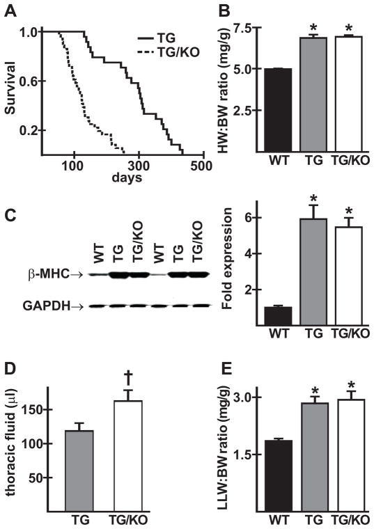
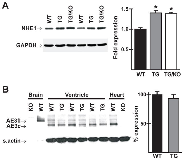
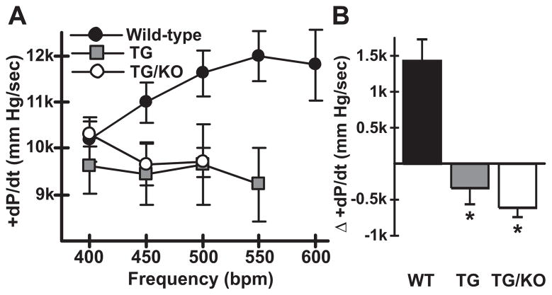
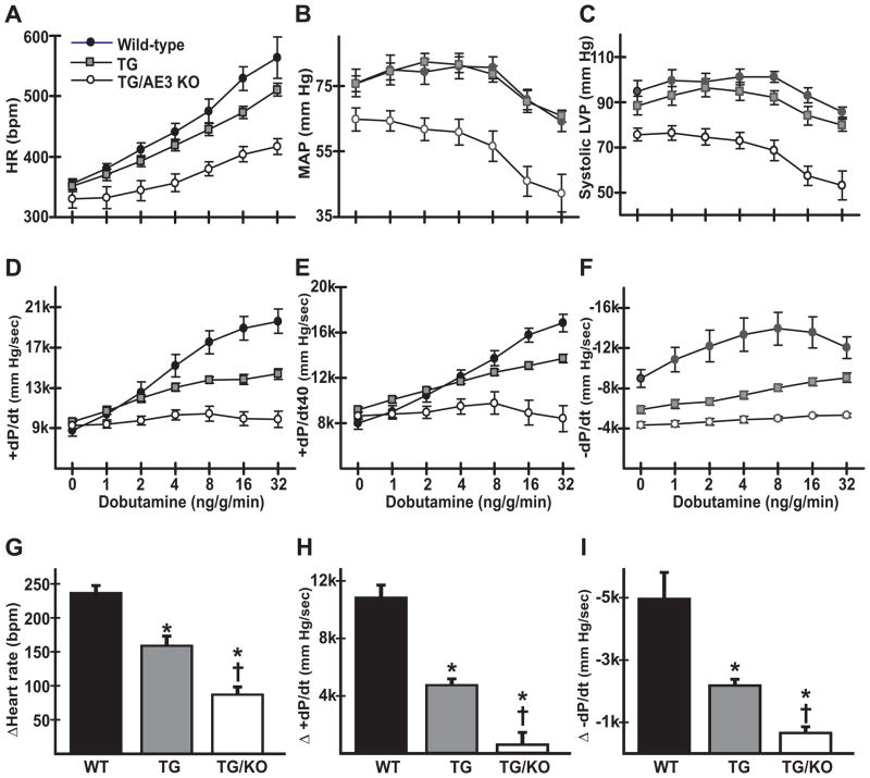
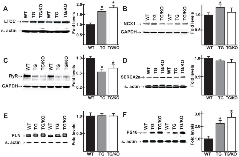
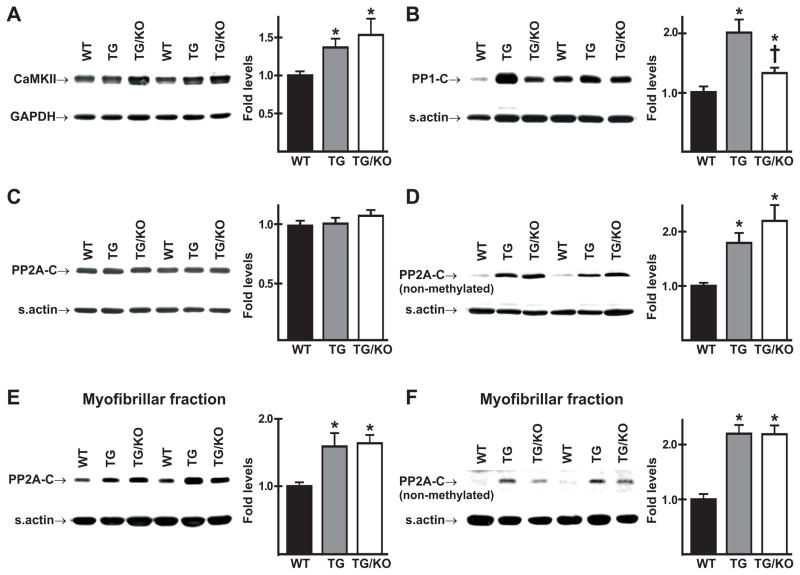
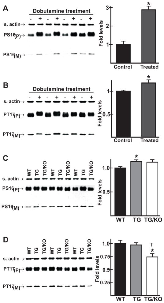
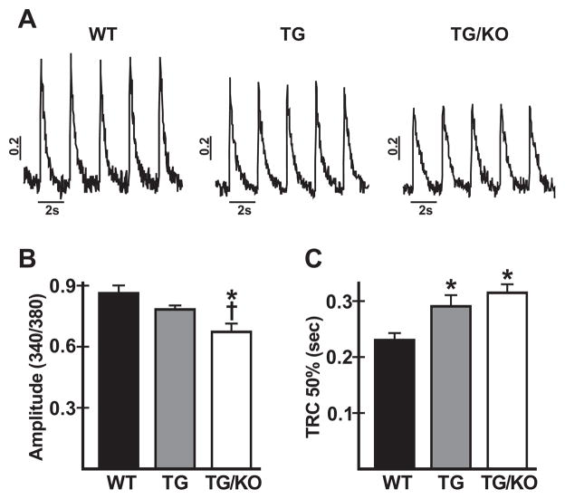
The AE3 Cl(-)/HCO(3)(-) exchanger is abundantly expressed in the sarcolemma of cardiomyocytes, where it mediates Cl(-)-uptake and HCO(3)(-)-extrusion. Inhibition of AE3-mediated Cl(-)/HCO(3)(-) exchange has been suggested to protect against cardiac hypertrophy; however, other studies indicate that AE3 might be necessary for optimal cardiac function. To test these hypotheses we crossed AE3-null mice, which appear phenotypically normal, with a hypertrophic cardiomyopathy mouse model carrying a Glu180Gly mutation in α-tropomyosin (TM180). Loss of AE3 had no effect on hypertrophy; however, survival of TM180/AE3 double mutants was sharply reduced compared with TM180 single mutants. Analysis of cardiac performance revealed impaired cardiac function in TM180 and TM180/AE3 mutants. TM180/AE3 double mutants were more severely affected and exhibited little response to β-adrenergic stimulation, a likely consequence of their more rapid progression to heart failure. Increased expression of calmodulin-dependent kinase II and protein phosphatase 1 and differences in methylation and localization of protein phosphatase 2A were observed, but were similar in single and double mutants. Phosphorylation of phospholamban on Ser16 was sharply increased in both single and double mutants relative to wild-type hearts under basal conditions, leading to reduced reserve capacity for β-adrenergic stimulation of phospholamban phosphorylation. Imaging analysis of isolated myocytes revealed reductions in amplitude and decay of Ca(2+) transients in both mutants, with greater reductions in TM180/AE3 mutants, consistent with the greater severity of their heart failure phenotype. Thus, in the TM180 cardiomyopathy model, loss of AE3 had no apparent anti-hypertrophic effect and led to more rapid decompensation and heart failure.


Figures

Fig. 1
Fig. 1
TM180/AE3 mutant mice exhibit reduced survival but no change in hypertrophy compared to TM180 single mutants. (A) Survival curves of transgenic TM180 mice (TG) and TG mice with AE3 knocked out (TG/KO); n = 26 TG and 38 TG/KO male mice; p < 0.0001 by Kaplan-Meier log-rank analysis. (B) Heart weight/body weight ratios for 2.5-month-old male and female wild-type (WT), TG, and TG/KO mice showed similar hypertrophy in TG (6.84 ± 0.02 mg/g) and TG/KO (6.91 ± 0.01) compared with WT (4.97 ± 0.04); n = 20 WT, 22 TG, and 23 TG/KO mice; *p < 0.001 vs WT. (C) Immunoblot analysis showed upregulation of β-myosin heavy chain (β-MHC) in ventricles of 2.5- or 3-month-old TG and TG/KO mice; n = 6 males of each genotype; *p < 0.01 vs WT. (D) Accumulation of thoracic fluid was higher in 2.5-month-old TG/KO than in TG mice; n = 17 TG and 14 TG/KO male and female mice; p < 0.05 vs TG. (E) Left lung weight/body weight ratios (LLW/BW in mg/g) were similarly elevated in 2.5-month-old male and female TG and TG/KO mice; n = 9 WT, 8 TG and 10 TG/KO; *p < 0.01 vs WT.
Fig. 2
Fig. 2
NHE1 and AE3 protein expression in ventricles of mutant and wild-type mice. (A) Immunoblot analysis revealed increased expression of NHE1 in ventricles of 3-month-old TM180 transgenic (TG) and TM180/AE3 double mutant (TG/KO) mice; n = 6 male mice of each genotype; *p < 0.001 vs WT. (B) Immunoblotting using an AE3 antibody that identifies both full length (AE3fl) and cardiac (AE3c) forms of AE3 revealed no significant change in ventricles of TM180 single mutant (TG) vs. WT male mice. Note high expression of AE3fl and AE3c in WT brain and whole heart, respectively, and absence of these variants in KO brain and whole heart.
Fig. 3
Fig. 3
TM180 (TG) and TM180/AE3 (TG/KO) mutants exhibit a negative force-frequency response. Hearts of anesthetized surgically-instrumented 3-month-old mice were subjected to atrial pacing beginning at 400 beats per minute (bpm) and contractile parameters were measured. n = 5 WT, 4 TG, and 5 TG/KO mice, with 2 males and either 2 or 3 females of each genotype. WT mice could be paced to 550 and 600 bpm but some TG and TG/KO mice could not. If fewer than 3 mice could achieve a given frequency, such as TG/KO at 550 bpm, the data were not plotted. (A) A positive FFR with respect to maximum +dP/dt (mm Hg/sec) was observed in WT mice but not in TG and TG/KO mice. (B) Difference in +dP/dt at 400 bpm and 500 bpm revealed a negative FFR in TG and TG/KO mice. *p < 0.02 vs WT.
Fig. 4
Fig. 4
β-adrenergic stimulation of cardiovascular performance is severely reduced in TM180/AE3 double mutants. Pressure measurements were recorded using transducers in the left ventricle and right femoral artery of anesthetized 2.5-month-old WT, TM180 (TG), or TM180/AE3 (TG/KO) mice of each genotype under basal conditions and in response to β-adrenergic stimulation (intravenous infusion of increasing doses of dobutamine). Heart rate (A), mean arterial pressure (B), systolic left ventricular pressure (C), maximum +dP/dt in mm Hg/sec (D), +dP/dt at 40 mm Hg (E), and minimum −dP/dt in mm Hg/sec (F) are shown for WT, TG, and TG/KO mice. Differences between basal and maximum values during β-adrenergic stimulation are shown for heart rate (G), +dP/dt (H), and -dP/dt (I). n = 7 WT (4 female, 3 male), 9 TG (5 female, 4 male), and 6 TG/KO (4 female, 2 male) mice. *p < 0.05 vs WT, p < 0.05 vs TG.
Fig. 5
Fig. 5
Expression of proteins involved in Ca2+ handling. Immunoblot analysis was performed using homogenates of ventricles from 2.5- or 3-month-old male WT, TM180 (TG), and TM180/AE3 (TG/KO) mice. Representative immunoblots and relative expression levels are shown for the L-type Ca channel, (A), NCX1 Na+/Ca2+ exchanger (B), ryanodine receptor (C), SERCA2a Ca2+ pump (D), phospholamban (E), and PLN phosphorylated on Ser16 (PS16) (F). n = at least 6 mice of each genotype, except for panel E, in which n = 3 of each genotype; *p < 0.05 vs WT.
Fig. 6
Fig. 6
Expression of Ca2+-calmodulin-dependent protein kinase II (CaMKII) and catalytic subunits of protein phosphatases PP1 and PP2A (PP1-C and PP2A-C). Immunoblot analysis was performed using homogenates of ventricles (A-D) or myofibrillar fractions (E,F) from 2.5-month-old male WT, TM180 (TG), and TM180/AE3 (TG/KO) mice. Representative immunoblots and relative expression levels are shown for (A) CamKII, (B) PP1-C, (C) total PP2A-C (non-methylated and methylated), (D) non-methylated form of PP2A-C, (E) total (non-methylated and methylated) PP2A-C associated with the myofribrillar fraction, (F) non-methylated PP2A-C associated with the myofribrillar fraction. n = at least 6 male mice of each genotype. *p < 0.05 vs WT; p < 0.05 vs TG.
Fig. 7
Fig. 7
Effects of β-adrenergic stimulation in vivo on phosphorylation of phospholamban in WT and mutant mice. Mice were anesthetized and surgically-instrumented as in Fig. 3. Ventricles were collected under basal conditions or after maximum stimulation with dobutamine, and immunoblot analysis of homogenates was performed using antibodies that recognize phosphoserine 16 (PS16) or phosphothreonine 17 (PT17) of PLN, with P and M designating pentameric and monomeric forms. (A) PS16 and (B) PT17 levels in WT ventricles under both basal (− dobutamine) and stimulated (+ dobutamine) conditions; *p < 0.05 vs control. (C) PS16 and (D) PT17 levels in WT, TM180 (TG), and TM180/AE3 (TG/KO) ventricles following maximum β-adrenergic stimulation; *p < 0.05 vs WT; p < 0.05 vs TG. n = 4 (A and B) or 3 (C and D) 2.5-month-old male mice of each genotype.
Fig. 8
Fig. 8
Analysis of Ca2+ transients in cardiac myocytes from WT and mutant mice. Myocytes were isolated from 3-month-old WT, TM180 (TG), and TM180/AE3 (TG/KO) mice and loaded with Fura-2AM. Ca2+ transients were analyzed during stimulation at 0.5 Hz. (A) Representative tracings of Ca2+ transients for the three genotypes, determined as fluorescence ratios at 340/380 nm. (B) Amplitudes of Ca2+ transients (systolic - diastolic values) for all three genotypes. (C) Time to 50% recovery of the Ca2+ transient (TRC 50%) for all three genotypes. For all three genotypes, n = 6 mice (4 females and 2 males) with 9–16 cells for each mouse. *p < 0.03 vs WT; p < 0.05 vs TG.

Similar articles

Cited by

References

    1. Linn SC, Kudrycki KE, Shull GE. The predicted translation product of a cardiac AE3 mRNA contains an N terminus distinct from that of the brain AE3 Cl−/HCO3− exchanger. Cloning of a cardiac AE3 cDNA, organization of the AE3 gene, and identification of an alternative transcription initiation site. J Biol Chem. 1992;267:7927–35. - PubMed
    1. Yannoukakos D, Stuart-Tilley A, Fernandez HA, Fey P, Duyk G, Alper SL. Molecular cloning, expression, and chromosomal localization of two isoforms of the AE3 anion exchanger from human heart. Circ Res. 1994;75:603–14. - PubMed
    1. Linn SC, Askew GR, Menon AG, Shull GE. Conservation of an AE3 Cl−/HCO3− exchanger cardiac-specific exon and promoter region and AE3 mRNA expression patterns in murine and human hearts. Circ Res. 1995;76:584–91. - PubMed
    1. Alvarez BV, Kieller DM, Quon AL, Markovich D, Casey JR. Slc26a6: a cardiac chloride-hydroxyl exchanger and predominant chloride-bicarbonate exchanger of the mouse heart. J Physiol. 2004;561:721–34. - PMC - PubMed
    1. Niederer SA, Swietach P, Wilson DA, Smith NP, Vaughan-Jones RD. Measuring and modeling chloride-hydroxyl exchange in the guinea-pig ventricular myocyte. Biophys J. 2008;94:2385–403. - PMC - PubMed

Publication types

MeSH terms