. 2011 Jan 14;405(2):410-26.
doi: 10.1016/j.jmb.2010.10.040. Epub 2010 Nov 5.

Her2-specific multivalent adapters confer designed tropism to adenovirus for gene targeting

Affiliations

Her2-specific multivalent adapters confer designed tropism to adenovirus for gene targeting

Birgit Dreier et al. J Mol Biol. .

Abstract

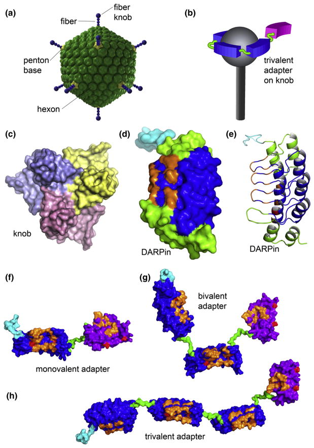
Adenoviruses (Ads) hold great promise as gene vectors for diagnostic or therapeutic applications. The native tropism of Ads must be modified to achieve disease site-specific gene delivery by Ad vectors and this should be done in a programmable way and with technology that can realistically be scaled up. To this end, we applied the technologies of designed ankyrin repeat proteins (DARPins) and ribosome display to develop a DARPin that binds the knob domain of the Ad fiber protein with low nanomolar affinity (K(D) 1.35 nM) and fused this protein with a DARPin specific for Her2, an established cell-surface biomarker of human cancers. The stability of the complex formed by this bispecific targeting adapter and the Ad virion resulted in insufficient gene transfer and was subsequently improved by increasing the valency of adapter-virus binding. In particular, we designed adapters that chelated the knob in a bivalent or trivalent fashion and showed that the efficacy of gene transfer by the adapter-Ad complex increased with the functional affinity of these molecules. This enabled efficient transduction at low stoichiometric adapter-to-fiber ratios. We confirmed the Her2 specificity of this transduction and its dependence on the Her2-binding DARPin component of the adapters. Even the adapter molecules with four fused DARPins could be produced and purified from Escherichia coli at very high levels. In principle, DARPins can be generated against any target and this adapter approach provides a versatile strategy for developing a broad range of disease-specific gene vectors.


Figures

Fig. 1
Fig. 1
The structures of Ad5 and its fiber knob domain, monovalent DARPin, and multivalent DARPin-derived adapters. (a) The icosahedral shell of the Ad5 virion is formed primarily by the hexon protein. Each of the 12 vertices contains the penton capsomer that consists of a trimeric fiber protein anchored within the pentameric penton base. The knob domain located at the end of the fiber binds the primary receptor. (b) The illustration shows the intended arrangement, with the virus fiber protein (gray) bound by three adapter DARPins (blue) connected to a Her2-binding DARPin (magenta) in one polypeptide chain. Please note (c)–(e) and (f)–(h) are drawn to scale. (c) Trimeric knob protein, viewed along the 3-fold symmetry axis from the side distal to the virion surface (PDB ID 1knb). Individual subunits are shown in different colors. (d) and (e) Monovalent DARPin 2E6 model showing the N-terminal His6 tag (cyan), capping repeats (green), and consensus repeats (blue) with seven randomized residues (orange) per repeat for target binding in either space-filling (d) or ribbon representation (e). Each 33 amino acid repeat in the DARPin consists of two antiparallel α-helices and a β-hairpin, resulting in a groove-like binding surface. The capping repeats provide a hydrophilic surface. The resulting DARPins consist of one contiguous polypeptide chain with a randomized binding surface. Bispecific adapter DARPins with varying valency are shown in (f) 2E6-G5, (g) (2E6)2-G5 and (h) (2E6)3-G5. The anti-knob DARPin 2E6 is shown with a blue surface; the anti-HER2 DARPin G5 is shown with a magenta surface, with the randomized residues in orange, the framework mutations in red, the N-terminal His6 tag in cyan and the (Gly4Ser)n linkers in green. The linkers shown between the 2E6 DARPins are (Gly4Ser)2, and (Gly4Ser)3 between 2E6 and G5.
Fig. 2
Fig. 2
Binding of knob-specific DARPins to virion-associated fibers. DARPins F4 (filled bars) and 2E6 (open bars), immobilized via their biotinylated AviTag to a streptavidin-coated ELISA plate, were probed with the particles of Ad vectors (Ad5lucΔF, fiber-depleted virus; Ad5Luc1, virus containing the wild-type knob; Ad5LucFΔ-TAYT, virus containing the knob with a 4 bp deletion in the FG loop of the knob) shown below the graph. DARPin-bound Ads were detected with an anti-Ad antibody and HRP-labeled secondary antibody. Background determined in casein-coated wells was at an absorbance at 490 nm (A490) of 0.2 and has not been subtracted. Error bars show standard deviation calculated for triplicate data points.
Fig. 3
Fig. 3
Adapter-mediated transduction of cultured human cells. Her2-negative 293 cells and Her2-expressing stably transfected 293/Her2 cells were infected with Ad5LucFΔTAYT alone (shown as virus) or preincubated with the adapter fusion proteins (shown below the graph) at adapter-to-fiber ratios of 1, 10 or 100. The virus was added to cells at a multiplicity of infection of 100 VP/cell. Graphs show the activity of luciferase reporter in the lysates of infected cells in relative light units. Error bars show standard deviations calculated for triplicate data points.
Fig. 4
Fig. 4
Determination of stoichiometry of DARPin binding to the knob by SEC. (a)–(d) Profiles of mixtures of 2E6 DARPin with the knob protein (one, two, three or four DARPin molar equivalents added per trimeric knob) resolved by SEC. Ratios at which the proteins were mixed are shown in the upper right corner of each panel. Profiles of elution for the knob, DARPin and DARPin-knob mixtures are shown by the dotted, broken and unbroken lines, respectively. (e) Overlay of the curves corresponding to the DARPin-knob mixtures (as in (a)–(d)). (f) SDS-PAGE of mixtures of 2E6 with the knob at indicated ratios, which were used to create a calibration curve to measure the ratio of the proteins in the chromatography peak containing the DARPin–knob complexes. (g) SEC-MALS of DARPin–knob mixtures. The knobΔTAYT protein at a concentration of 5 μM was incubated with different concentrations of DARPin 2E6 (3.75, 11.25, 22.5 and 90 μM). Complex formation was analyzed by size-exclusion chromatography coupled to MALS. The curves shown represent the absorption at 280 nm (peaks; left-hand y-axis). The determined mass of the knobΔTAYT trimer, DARPin 2E6 and the complex are given on top of the corresponding protein peaks (right-hand y-axis). (h) ITC of knobΔTAYT protein (34 μM) with DARPin 2E6 (770 μM stock). Top panel, raw instrumental output (differential power as a function of time). Bottom panel, binding isotherm. The heats measured at each titration step were normalized for the molar concentration and corrected for unspecific heats (symbols). The unbroken line is the best fit according to a binding model assuming identical and independent binding sites. The best fitting parameters are: molar enthalpy (ΔH), −26.5±0.9 kcal mol−1; and number of binding sites (n), 2.5±0.2. Note that the concentrations are so far above KD that a reliable determination of KD is not possible.
Fig. 5
Fig. 5
Optimization of gene transfer. (a) Bivalent targeting adapters enable improved Ad transduction of Her2-expressing cells. V, virus alone; MVA, monovalent 2E6-G5 adapter, virus in complex with the control monovalent adapter 2E6-G5; L1-L6, virus in complex with one of the six bivalent and bispecific adapters (2E6-Ln-2E6-L3-G5) each containing two copies of 2E6 connected with linkers of various length. L1 denotes Gly4Ser, L2 denotes (Gly4Ser)2, etc. Adapter-to-fiber ratios are shown below the graph. (b) Increased valency of targeting adapter yields improved transduction of Her2-expressing cells. Transgene expression by the vector alone (V) or by the vector in a complex with mono- (1), bi- (2) or trivalent (3) adapters in which the 2E6 DARPin modules are spaced by a (Gly4Ser)2 linker. Transgene expression by CAR binding-ablated Ad in cultured cells is shown as activity of luciferase reporter in the lysates of infected cells displayed in relative light units. Error bars show standard deviations calculated for triplicate data points.
Fig. 6
Fig. 6
Functional affinity of the monovalent, bivalent and trivalent adapters. Competition experiments in solution were performed using SPR technology. Constant concentrations of the corresponding adapter (14–18 nM) were first incubated with increasing amounts of knobΔ-TAYT in solution. Then the binding of free adapter to the knobΔTAYT immobilized on a biosensor chip was measured. The initial on-rates plotted against the total soluble knob subunit concentration and the corresponding fits are shown for the monovalent 2E6-G5 (a), bivalent (2E6)2-G5 (b) and trivalent (2E6)3-G5 (c). In each panel the data obtained in two replicate experiments are depicted with open and filled symbols. The dotted line in (b) and (c) is the fit of the monovalent adapter copied from (a).
Fig. 7
Fig. 7
Her2-binding G5 ligand is essential to the specificity of adapter-mediated transduction. Shown is the activity of luciferase reporter in relative light units in the lysates of cells transduced with virus alone (V) or in complex with either the (2E6)2-G5 (1) or its truncated analog (2E6)2-L (2) that lacks the G5 ligand. In addition, cells were preincubated with no inhibitor, free G5, or a free unrelated DARPin (E2-5), as shown below the graph. Error bars show standard deviations calculated for triplicate data points.

Similar articles

Cited by

References

    1. Amalfitano A, Parks RJ. Separating fact from fiction: assessing the potential of modified adenovirus vectors for use in human gene therapy. Curr Gene Ther. 2002;2:111–133. - PubMed
    1. Barouch DH, Pau MG, Custers JH, Koudstaal W, Kostense S, Havenga MJ, et al. Immunogenicity of recombinant adenovirus serotype 35 vaccine in the presence of pre-existing anti-Ad5 immunity. J Immunol. 2004;172:6290–6297. - PubMed
    1. Kobinger GP, Feldmann H, Zhi Y, Schumer G, Gao G, Feldmann F, et al. Chimpanzee adenovirus vaccine protects against Zaire Ebola virus. Virology. 2006;346:394–401. - PubMed
    1. Sullivan NJ, Geisbert TW, Geisbert JB, Xu L, Yang ZY, Roederer M, et al. Accelerated vaccination for Ebola virus haemorrhagic fever in non-human primates. Nature. 2003;424:681–684. - PMC - PubMed
    1. Burton JB, Johnson M, Sato M, Koh SB, Mulholland DJ, Stout D, et al. Adenovirus-mediated gene expression imaging to directly detect sentinel lymph node metastasis of prostate cancer. Nat Med. 2008;14:882–888. - PMC - PubMed

Publication types