Case Reports
. 2011 Jan 20;409(2):141-7.
doi: 10.1016/j.virol.2010.10.020. Epub 2010 Nov 5.

Computational analysis and identification of an emergent human adenovirus pathogen implicated in a respiratory fatality

Affiliations
Case Reports

Computational analysis and identification of an emergent human adenovirus pathogen implicated in a respiratory fatality

Christopher M Robinson et al. Virology. .

Abstract

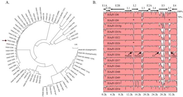
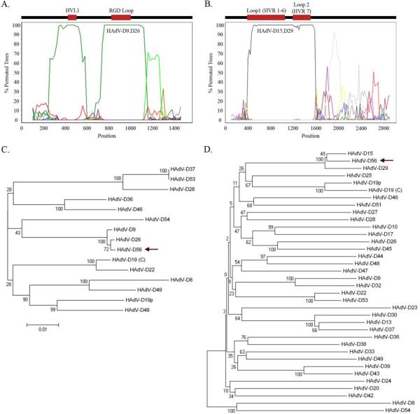
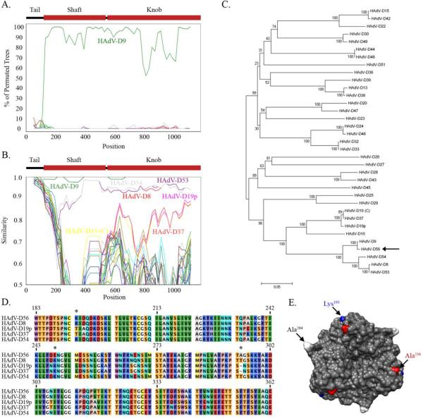
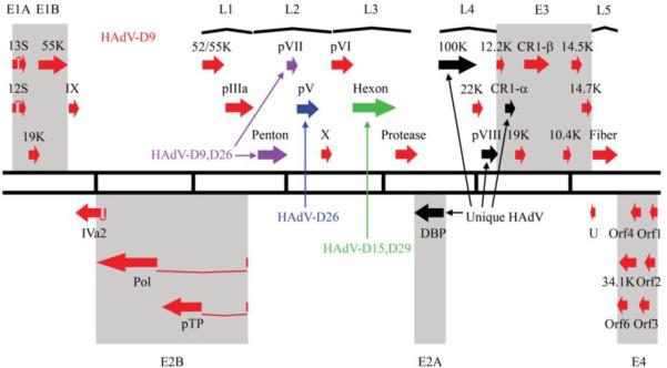
Adenoviral infections are typically acute, self-limiting, and not associated with death. However, we present the genomic and bioinformatics analysis of a novel recombinant human adenovirus (HAdV-D56) isolated in France that caused a rare neonatal fatality, and keratoconjunctivitis in three health care workers who cared for the neonate. Whole genome alignments revealed the expected diversity in the penton base, hexon, E3, and fiber coding regions, and provided evidence for extensive recombination. Bootscan analysis confirmed recombination between HAdV-D9, HAdV-D26, HAdV-D15, and HAdV-D29 in the penton base and hexon proteins, centered around hypervariable loops within the putative proteins. Protein structure analysis of the fiber coding region revealed similarity with HAdV-D8, HAdV-D9, and HAdV-D53, possibly accounting for the ocular tropism of the virus. Based on these data, this virus appears to be a new HAdV-D type (HAdV-D56), underscoring the importance of recombination events in human adenovirus evolution and the emergence of new adenovirus pathogens.


Figures

Fig. 1
Fig. 1
Whole genome analysis of HAdV-D56. (A) Bootstrap- confirmed neighbor-joining tree designed from MEGA 4.0.2 demonstrates relationships between HAdV-D56 and all other completely sequenced adenovirus genomes. (arrow: HAdV-D56). Bootstrap values for the species nodes were 100. The bootstrap value for the HAdV-D56 and HAdV-D9 node was also 100. (B) Global pairwise sequence comparison of HAdV-D56 with fourteen other completely sequenced HAdV-D types. Percent sequence conservation is reflected in the height of each data point along the y-axis. The penton base, hexon, E3, and fiber protein coding regions are divergent within species D, except for the penton base and fiber regions of HAdV-D9 and HAdV-D26 (*).
Fig. 2
Fig. 2
Bootscan recombination and phylogenetic analysis of HAdV-D56 penton base and hexon genes. (A) Bootscan analysis comparing the HAdV-D56 penton base gene with completely sequenced HAdV types. (B) Bootscan analysis comparing the HAdV-D56 hexon gene with other HAdV-D types. (C) Phylogenetic analysis of the penton base gene from completely sequenced HAdV-D types (arrow: HAdV-D56). (D) Phylogenetic analysis of the hexon gene from all available HAdV-D types (arrow: HAdV-D56). Bootstrap values below 80 are indicative of low confidence.
Fig. 3
Fig. 3
Genomic and structural analysis of the HAdV-D56 fiber. (A) Bootscan analysis comparing the HAdV-D56 fiber gene with each HAdV-D type. (B) Simplot analysis comparing the HAdV-D56 fiber gene with each HAdV-D type. (C) Phylogenetic analysis of sequenced HAdV-D fiber genes (arrow: HAdV-D56). (D) Multi-sequence alignment of the fiber knob designed from MEGA 4.0.2 software. HAdV-D56 unique residues compared to other EKC viruses designated by *. HAdV-D19(C) and HAdV-D53 were left out of the analysis based on 100% identity to HAdV-D37 and HAdV-D8, respectively. (E) HAdV-D56 protein trimer model based on homology modeling from the crystal structure of HAdV-D19p. Unique residues Lys193 and Ala294 are highlighted in blue and white, respectively. Ala236, which may play a key role in host receptor binding, is highlighted in red.
Fig. 4
Fig. 4
Transcriptional map of the recombinant HAdV-D56 genome. Genes with high sequence identity to HAdV-D9; HAdV-D9,D26; HAdV-D26; and HAdV-D15,D29 are indentified in red, purple, blue, and green, respectively, where HAdV-D9,D26 refers to genomic sequence for which HAdV-D9 and HAdV-D26 are nearly identical, and HAdVD15,D29 refers to sequence where HAdV-D15 and HAdV-D29 are nearly identical. Genes with unique sequence are identified in black.

References

    1. Abbondanzo SL, English CK, Kagan E, McPherson RA. Fatal adenovirus pneumonia in a newborn identified by electron microscopy and in situ hybridization. Arch Pathol Lab Med. 1989;113(12):1349–53. - PubMed
    1. Abzug MJ, Levin MJ. Neonatal adenovirus infection: four patients and review of the literature. Pediatrics. 1991;87(6):890–6. - PubMed
    1. Adrian T, Bastian B, Benoist W, Hierholzer JC, Wigand R. Characterization of adenovirus 15/H9 intermediate strains. Intervirology. 1985;23(1):15–22. - PubMed
    1. Arnberg N, Kidd AH, Edlund K, Nilsson J, Pring-Akerblom P, Wadell G. Adenovirus type 37 binds to cell surface sialic acid through a charge-dependent interaction. Virology. 2002;302(1):33–43. - PubMed
    1. Arnberg N, Mei Y, Wadell G. Fiber genes of adenoviruses with tropism for the eye and the genital tract. Virology. 1997;227(1):239–44. - PubMed

Publication types

MeSH terms

Associated data