. 2011 Feb 11;286(6):4659-69.
doi: 10.1074/jbc.M110.175638. Epub 2010 Nov 5.

The oligomeric state and arrangement of the active bacterial translocon

Affiliations

The oligomeric state and arrangement of the active bacterial translocon

Karine Deville et al. J Biol Chem. .

Abstract

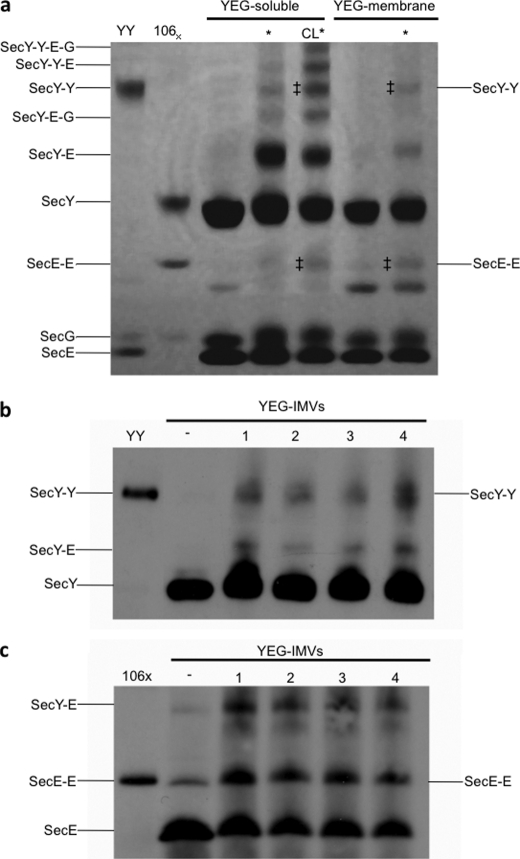
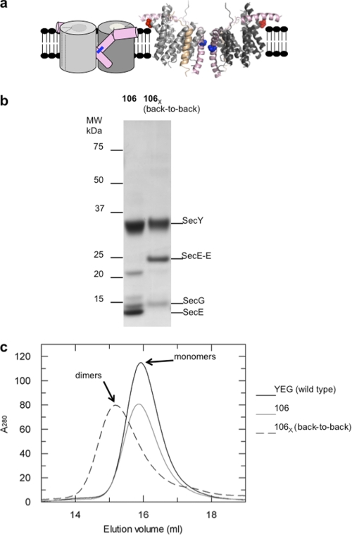
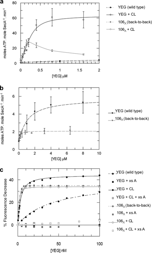
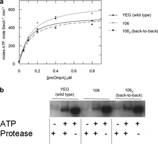
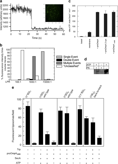
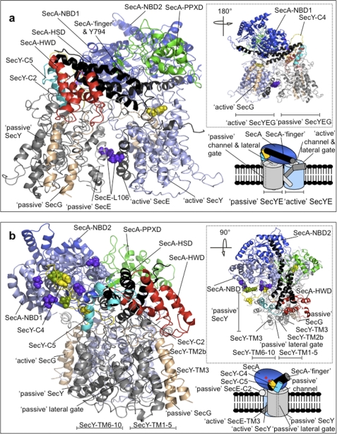
Protein secretion in bacteria is driven through the ubiquitous SecYEG complex by the ATPase SecA. The structure of SecYEG alone or as a complex with SecA in detergent reveal a monomeric heterotrimer enclosing a central protein channel, yet in membranes it is dimeric. We have addressed the functional significance of the oligomeric status of SecYEG in protein translocation using single molecule and ensemble methods. The results show that while monomers are sufficient for the SecA- and ATP-dependent association of SecYEG with pre-protein, active transport requires SecYEG dimers arranged in the back-to-back conformation. Molecular modeling of this dimeric structure, in conjunction with the new functional data, provides a rationale for the presence of both active and passive copies of SecYEG in the functional translocon.


Figures

FIGURE 1.
FIGURE 1.
Probing the oligomeric state of SecYEG in active reconstituted and native membranes. a, photo-cross-linking of detergent-solubilized SecYEG (YEG-soluble) in the absence and presence of 60 μm CL, or in reconstituted proteoliposomes (YEG-membrane), was performed by exposure to light radiation. Proteins were subsequently separated by SDS-PAGE and visualized by Coomassie Blue staining. A marker for cross-linked SecY-Y was provided by a genetically-fused, tandem dimer of SecY (YY) (13) and for SecE-E by SecYEL106CG (106X) (26). * denotes samples that have been irradiated. The predicted compositions indicated for the cross-linked species are consistent with their observed mobilities, and the bands indicated by ‡ were confirmed by mass spectrometry to contain accordingly SecY or SecE. b and c, IMVs overexpressing SecYEG (YEG-IMVs) were photo-cross-linked, separated by SDS-PAGE, transferred to nitrocellulose for Western blotting and probed with monoclonal antibodies against SecY (b) and SecE (c). − denotes no irradiation; samples in lanes 1 and 2 were irradiated for 90 s with 1 mm or 2 mm cross-linker, respectively, and likewise, lanes 3 and 4 for 180 s with 1 mm or 2 mm.
FIGURE 2.
FIGURE 2.
The back-to-back form of the SecYEG cross-linked in the membrane is stable and dimeric in solution. a, schematic side view and ribbon representation of the back-to-back (6) dimer of SecYEG: SecY subunits (dark and light gray), SecE (TMS 3 and the amphipathic cytosolic helix, pink), SecG (tan). In the scheme, SecG is not shown, and the channel and lateral gate (for TMS insertion (3), dashed lines) are closed. The cross-link between the two cysteines is shown by a blue bar. In the ribbon diagram, the cysteine residues employed for cross-linking, SecEL106C (26) (blue), and for labeling with AF488, SecYA103C (red; see Fig. 5), are shown in space-fill. b, non-reducing SDS-PAGE and c, size-exclusion chromatography analysis of the purified wild-type complex (YEG), and uncross-linked (106) and cross-linked (106X) forms of SecYEL106CG, the latter as a result of CuPh treatment of membranes prior to extraction and purification. The mobilities of the individual subunits and the cross-linked dimers of SecE (SecE-E) are indicated.
FIGURE 3.
FIGURE 3.
The back-to-back version of the SecYEG dimer provides a high affinity platform required for the productive association of SecA. a, analysis of SecA ATPase stimulation by monomeric (YEG) and back-to-back dimeric forms (106X) of SecYEG. The data obtained in the absence of CL are shown again, using an expanded scale in b. ATPase activity of SecA (0.3 μm) was measured in detergent solution (0.03% C12E9) in the presence of 40 μm CL, with increasing concentrations of either wild-type SecYEG (YEG, black solid line, open circles) or SecYEL106CG (106X, gray solid line, open circles), or in the absence of CL (black and gray dashed lines and filled circles, respectively). With the exception of SecYEL106CG +CL (106X + CL), which showed decreased activities at high concentrations, probably as the result of aggregation, the data were fitted according to a tight binding equation (“Experimental Procedures”). Error bars represent S.D. (n > 3). c, estimation of the affinity of the monomeric (YEG) and back-to-back dimeric forms (106X) of SecYEG for SecA by fluorescence spectroscopy. Wild-type SecYEG (YEG) or cross-linked SecYEL106CG (106X) were titrated into a solution of 2 nm SecA795Fl in the presence or absence of 40 μm CL. The data were fitted to Equations 2 or 3 (“Experimental Procedures”). Controls were performed with the addition of an excess of unlabeled wild-type SecA (xs A), which competed for the binding and obliterated the fluorescence change. Kd values from the fitting procedures are shown in Table 1.
FIGURE 4.
FIGURE 4.
The SecYEG complex held in the back-to-back orientation is fully active in ATP driven translocation. a, steady-state analysis of the translocation-coupled ATPase activity stimulated by membrane-bound SecYEG and proOmpA. ATPase activity of SecA (50 nm) was measured in the presence of 1 mm ATP and 0.6 μm SecYEG in proteoliposomes with increasing concentrations of proOmpA as indicated, applied from a stock dissolved in 6 m urea. Proteoliposomes were reconstituted with wild-type SecYEG (YEG), SecYEL106CG (106), or cross-linked SecYEL106CG (back-to-back dimer, 106X). The data sets were fitted according to Equation 1 (“Experimental Procedures”) and the parameters (Kd and kcat,) are shown in Table 2. b, measurement of in vitro translocation of proOmpA (0.7 μm) into the same SecYEG proteoliposomes (YEG, 106, and 106X) in the presence of SecA. The extent of proOmpA translocation, i.e. protection from protease digestion, was detected by Western blotting and compared with a measure of 10% of the total proOmpA in each reaction (ATP +, Protease −) and a negative control performed in the absence of ATP (ATP −, Protease +).
FIGURE 5.
FIGURE 5.
Single molecule fluorescence measurements of protein translocation and the oligomeric state of SecYEG. a, representative time-course of fluorescence intensity showing quantized photobleaching in a single step for a vesicle harboring a single SecY103-AF488EG molecule. The images were recorded at a video rate of 60 ms/image frame with an Andor iXon EMCCD camera. The y axis plots the photon counts (not corrected for background). Inset, representative image obtained by TIRF microscopy showing a field of tethered SecY103-AF488EG vesicles. b, distribution of different types of photobleaching events observed for 500 vesicles reconstituted at different LPRs. Trajectories that could not be clearly described as representing single, double or discrete numbers of multiple step(s) are designated “unclassified”. c, ATPase activity of 0.3 μm SecA measured in the presence of 1 mm ATP in the absence (basal) and in the presence of 1 μm SecYEG in proteoliposomes without protein substrate (membrane) or with 0.7 μm of either wild-type proOmpA, proOmpAΔ176 (proOmpA 2 cys) or Alexa Fluor 488-labeled proOmpAΔ176 (proOmpAA488). Error bars represent S.D. (n = 3). d, in vitro translocation reactions carried out with 1 μm SecYEG in proteoliposomes, 50 nm SecA, 1 mm ATP, and 0.7 μm of labeled proOmpAΔ176 (proOmpAA488). Successfully translocated proOmpA substrate was protected from proteinase K (PK) digestion and detected using a Typhoon 9400 fluorescence imager. e, comparison of the numbers of fluorescent vesicles observed following addition to proteoliposomes, harboring different forms of SecYEG, with SecA and proOmpAA488, in the presence or absence of ATP, and subsequent treatment with tryptophan. Results are shown as mean ± S.D. (n = 5). The vesicles tested contained either single copies of SecY103-AF488EG ((Y*EG)1; LPR = 73,000:1), unlabeled single copies of SecYEG monomers ((YEG)1 wild-type; LPR = 73,000:1), unlabeled multiple copies of SecYEG ((YEG)n wild-type; LPR = 730:1), labeled tandem SecY dimers ((YY*EG)1; LPR 73,000:1), or single copies of unlabeled SecYEL106CG cross-linked in the back-to-back configuration ((106X)1 back-to-back; LPR = 73,000:1).
FIGURE 6.
FIGURE 6.
Atomic model of the E. coli membrane-bound translocon SecA-(SecYEG)2. a, cartoon representation of face-on and b, end-on side-views of dimeric SecYEG associated with SecA. The insets in the lower right are schematic representations of the model views in the back-to-back configuration, where the active and passive SecYEG protomers in the dimer are drawn with the channels respectively partially open (white and solid line ellipsoid) and closed (gray and dashed line ellipsoid); in this arrangement, the lateral gates (dashed gray lines) face away from each other. In the model, the passive and active SecY copies are colored in light gray and light blue, respectively. SecE is represented in light and dark gray for the passive and active complex, respectively. SecG is colored in tan. Similarly, differential coloring is used to highlight the individual domains of SecA: NBD1 (light blue), NBD2 (blue), HSD including the two-helix finger (black), PPXD (green), and HWD (red). The cytoplasmic loops of the passive SecY between TMS 6–7 (SecY-C4) and TMS 8–9 (SecY-C5) are shown respectively in yellow and cyan. a, SecE residue Leu-106 is shown in solid molecular representation in purple, while SecA residue Tyr-794 at the end of the two-helix finger is similarly shown in yellow. b, solid residues on NBD1 and SecY-C4 show the residues that could (purple and green), or could not (light blue and yellow) be cross-linked (14) (see supplemental Fig. S2, b and c for more detail). The insets at the top right of a and b show alternative views, rotated as indicated compared with the main figures.

Similar articles

Cited by

References

    1. Lill R., Cunningham K., Brundage L. A., Ito K., Oliver D., Wickner W. (1989) EMBO J. 8, 961–966 - PMC - PubMed
    1. Economou A., Pogliano J. A., Beckwith J., Oliver D. B., Wickner W. (1995) Cell 83, 1171–1181 - PubMed
    1. van den Berg B., Clemons W. M., Jr., Collinson I., Modis Y., Hartmann E., Harrison S. C., Rapoport T. A. (2004) Nature 427, 36–44 - PubMed
    1. Zimmer J., Nam Y., Rapoport T. A. (2008) Nature 455, 936–943 - PMC - PubMed
    1. Mitra K., Schaffitzel C., Shaikh T., Tama F., Jenni S., Brooks C. L., 3rd, Ban N., Frank J. (2005) Nature 438, 318–324 - PMC - PubMed

Publication types

MeSH terms