. 2010 Dec;177(6):3202-14.
doi: 10.2353/ajpath.2010.100594. Epub 2010 Nov 5.

Blockade of transforming growth factor-beta1 accelerates lymphatic regeneration during wound repair

Affiliations

Blockade of transforming growth factor-beta1 accelerates lymphatic regeneration during wound repair

Tomer Avraham et al. Am J Pathol. 2010 Dec.

Abstract

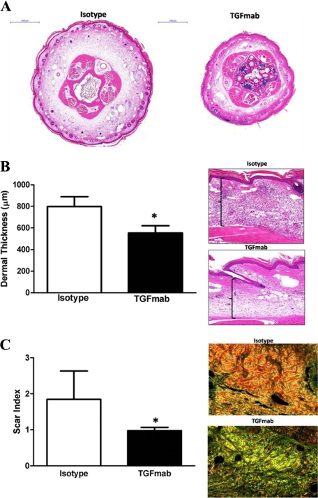
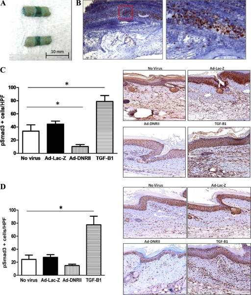
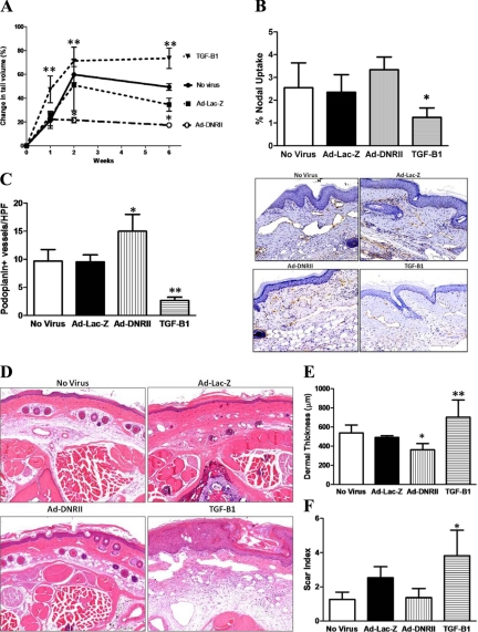
Lymphedema is a complication of cancer treatment occurring in approximately 50% of patients who undergo lymph node resection. Despite its prevalence, the etiology of this disorder remains unknown. In this study, we determined the effect of soft tissue fibrosis on lymphatic function and the role of transforming growth factor (TGF)-β1 in the regulation of this response. We determined TGF-β expression patterns in matched biopsy specimens collected from lymphedematous and normal limbs of patients with secondary lymphedema. To determine the role of TGF-β in regulating tissue fibrosis, we used a mouse model of lymphedema and inhibited TGF-β function either systemically with a monoclonal antibody or locally by using a soluble, defective TGF-β receptor. Lymphedematous tissue demonstrated a nearly threefold increase in the number of cells that stained for TGF-β1. TGF-β inhibition markedly decreased tissue fibrosis, increased lymphangiogenesis, and improved lymphatic function compared with controls. In addition, inhibition of TGF-β not only decreased TGF-β expression in lymphedematous tissues, but also diminished inflammation, migration of T-helper type 2 (Th2) cells, and expression of profibrotic Th2 cytokines. Similarly, systemic depletion of T-cells markedly decreased TGF-β expression in tail tissues. Inhibition of TGF-β function promoted lymphatic regeneration, decreased tissue fibrosis, decreased chronic inflammation and Th2 cell migration, and improved lymphatic function. The use of these strategies may represent a novel means of preventing lymphedema after lymph node resection.


Figures

Figure 1
Figure 1
Lymphedema is associated with increased expression of TGF-β1. A: Immunohistochemical analysis of matched skin biopsy specimens from lymphedematous and normal limbs of patients demonstrates increased intracellular and extracellular staining for TGF-β1 in lymphomatous tissues. Representative figures from two patients are shown at 40× magnification. B: Number of TGF-β1+ cells/High Power Field in lymphomatous and normal tissues (mean ± SD; *P < 0.04). C: Fold change in number of TGF-β1+ cells/HPF in lymphedematous versus normal limbs (mean ± SD; *P < 0.02).
Figure 2
Figure 2
Systemic TGF-β1 blockade decreases tail lymphedema and improves lymphatic regeneration. A: Tail volume measurements in isotype control and TGFmab-treated animals (mean ± SEM; *P < 0.05). B: Representative photographs of isotype- (left) and TGFmab (right)-treated animals. Note improved wound repair, minimal edema, and lack of tail curvature indicative of fibrosis in TGFmab-treated animals as compared with controls. C: Representative microlymphangiography demonstrates flow of fluorescent-tagged large molecular weight dextran across the wound only in the TGFmab-treated animals. In contrast, isotype controls demonstrate pooling of fluorescence distal to the wound. D: Tc99 lymphoscintigraphy demonstrating nearly ninefold greater lymphatic transport to lymph nodes at the base of the tail in TGFmab-treated mice as compared with isotype control (mean ± SEM; *P < 0.0001). Representative lymphoscintographs are presented (arrowhead: injection site near tip of tail; arrow: lymph node). E and F: Quantitation of podoplanin-positive lymphatic vessels demonstrates nearly twofold increase in number of vessels in tail tissues located distal to the zone of lymphatic disruption in mice treated with TGFmab as compared with isotype controls (mean ± SD; P < 0.0001). Representative 20× micrographs demonstrating scarce and dilated podoplanin positive vessels (arrow) in isotype-treated mice and abundant collapsed lymphatics (arrowheads) in TGFmab-treated mice. G and H: Quantitation of vWF-positive blood vessels (arrows), demonstrates a nonsignificant trend toward lower numbers of blood vessels in the TGFmab-treated animals.
Figure 3
Figure 3
Systemic TGF-β1 blockade decreases tissue fibrosis secondary to lymphatic fluid stasis. A: Representative cross-sectional histology of isotype- (left) and TGFmab-treated animals 6 weeks after surgery. Twofold magnification of both sections is shown. Note marked increase in ECM deposition, hypercellularity, and dilated lymphatics in isotype-treated animals. B: Dermal thickness measurement in isotype- and TGFmab-treated animals (mean ± SD; *P < 0.001). Representative figures of high-power (20×) views of longitudinal sections from isotype- (top) and TGFmab- (bottom) treated animals. C: Scar index calculation in isotype- and TGFmab-treated animals in tissues distal to the zone of lymphatic obstruction harvested 6 weeks after surgery (mean ± SD; *P < 0.01). Representative 20× micrographs demonstrate a predominantly red/orange birefringence (consistent with increased fibrosis) in isotype-treated animals (top). In contrast, TGFmab-treated animals demonstrate primarily a yellow/green birefringence, indicating normal ECM deposition.
Figure 4
Figure 4
Systemic TGF-β1 blockade reduces lymphedema induced chronic inflammation. A: Quantification of the number of CD45+ leukocytes/HPF in tissues located proximal or distal to the zone of lymphatic obstruction in isotype- (white bars) or TGFmab-treated animals (black bars; mean ± SD; *P < 0.001). Representative 20× micrographs are shown to the right. B: Quantification of CD4+ cells/HPF in tissues located proximal or distal to the zone of lymphatic obstruction in isotype- (white bars) or TGFmab-treated animals (black bars; mean ± SD; *P < 0.001). Representative 20× micrographs are shown to the right. Note gradients of inflammation with more CD4+ cells present distal to the zone of obstruction and reduction in cell number resulting from TGFmab treatment. C and D: Quantification of presumptive Th1 (IFN-γ+/CD4+) and Th2 (IL-13+/CD4+) cells in TGFmab- or isotype-treated animals 6 weeks after tail excision (mean ± SD; *P < 0.001). E: Representative Western blot analysis of tail soft tissues localized distal to the zone of lymphatic obstruction 6 weeks after surgery. Treatment with TGFmab resulted in marked reductions in expression of CD4, IL-4, IL-13, and TGF-β1. In contrast, only modest differences were noted in the expression of F4/80, VEGF-C, or VEGF-A. All experiments were performed in duplicate. F: Representative Western blot analysis of tissues located distal to the zone of lymphatic obstruction in isotype- or CD3-depleted mice. Again, note decreased expression of CD4, IL-4, IL-13, and TGF-β1. All experiments were performed in duplicate.
Figure 5
Figure 5
Adenoviruses can promote high-level transgene expression in mouse tail wound. A: A portion of tail transfected with Ad-Lac-Z was stained for β-galactosidase activity 1 week after surgery and transfect. The intense bluish staining, particularly at the wound, indicates increased activity and effective transfection. B: Immunohistochemistry for β-galactosidase demonstrated extensive expression in the peri-wound region 1 week after surgery and transfection with LacZ adenovirus. (Red Box: region of intense staining is magnified) C and D: Quantification of pSmad3+ cells/HPF in nonvirus, Ad-Lac-Z, Ad-DNRII, and recombinant TGF-β1 treated animals 1 (C) and 6 (D) weeks after surgery/transfection. Representative 40× images are shown to the right. Note significant decrease in the number of pSmad3+ cells/HPF in Ad-DNRII treated animals 1 week after surgery (mean ± SD; *P < 0.001). By 6 weeks, the only significant difference among groups was the number of PSmad-3 positive cells in the recombinant TGF-β1 treated group; *P < 0.001.
Figure 6
Figure 6
Local blockade of TGF-β1 activity decreases tail edema, increases lymphangiogenesis, and decreases tissue fibrosis. A: Tail volume measurements in nonvirus, Ad-Lac-Z, Ad-DNRII, and recombinant TGF-β1 treated animals at various time points after surgery (mean ± SD; **P < 0.01). B: Lymphoscintigraphy with Tc99 demonstrated modest, though nonsignificant, increase in nodal uptake in Ad-DNRII-treated animals. In contrasts, animals treated with recombinant TGF-β1 demonstrated markedly decreased lymphatic uptake (mean ± SEM; *P < 0.001). C: Quantification of the number of podoplanin+ lymphatic vessels in the various experimental groups 6 weeks after surgery. Mean ± SD; *P < 0.002; **P < 0.001. Representative 20× micrographs are shown to the right. D: Histological analysis of cross-sections of the tails demonstrating decreased ECM deposition and cellularity in Ad-DNRII-treated animals as compared with other groups. Representative 20× micrographs are shown. TGF-treated animals had greater swelling and lymphatic dilation compared with all other groups; 20× micrographs are presented. E: Quantitation of dermal thickness in distal tail sections 6 weeks after surgery. Treatment with Ad-DNRII decreased dermal thickness as compared with other groups. Mean ± SD; *P < 0.05. Treatment with recombinant TGF-β1 markedly increased dermal thickening; **P < 0.001. F: Quantitation of scar index by using Sirius red staining and computerized polarized light microscopy. Treatment with recombinant TGF-β1 significantly increased fibrosis; *P < 0.001. No significant differences were noted in the other groups.

References

    1. Velanovich V, Szymanski W. Quality of life of breast cancer patients with lymphedema. Am J Surg. 1999;177:184–187. discussion 188. - PubMed
    1. Beaulac SM, McNair LA, Scott TE, LaMorte WW, Kavanah MT. Lymphedema and quality of life in survivors of early-stage breast cancer. Arch Surg. 2002;137:1253–1257. - PubMed
    1. Purushotham AD, Upponi S, Klevesath MB, Bobrow L, Millar K, Myles JP, Duffy SW. Morbidity after sentinel lymph node biopsy in primary breast cancer: results from a randomized controlled trial. J Clin Oncol. 2005;23:4312–4321. - PubMed
    1. Tsai RJ, Dennis LK, Lynch CF, Snetselaar LG, Zamba GK, Scott-Conner C. The risk of developing arm lymphedema among breast cancer survivors: a meta-analysis of treatment factors. Ann Surg Oncol. 2009;16:1959–1972. - PubMed
    1. Koul R, Dufan T, Russell C, Guenther W, Nugent Z, Sun X, Cooke AL. Efficacy of complete decongestive therapy and manual lymphatic drainage on treatment-related lymphedema in breast cancer. Int J Radiat Oncol Biol Phys. 2007;67:841–846. - PubMed

Publication types

MeSH terms

Substances