. 2011 Jan;300(1):H388-96.
doi: 10.1152/ajpheart.00868.2010. Epub 2010 Nov 5.

Chronic cigarette smoking causes hypertension, increased oxidative stress, impaired NO bioavailability, endothelial dysfunction, and cardiac remodeling in mice

Affiliations

Chronic cigarette smoking causes hypertension, increased oxidative stress, impaired NO bioavailability, endothelial dysfunction, and cardiac remodeling in mice

M A Hassan Talukder et al. Am J Physiol Heart Circ Physiol. 2011 Jan.

Abstract

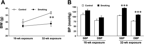
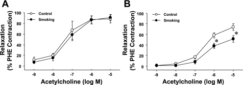
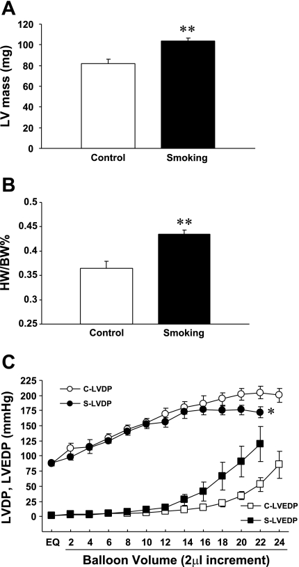
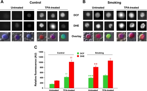
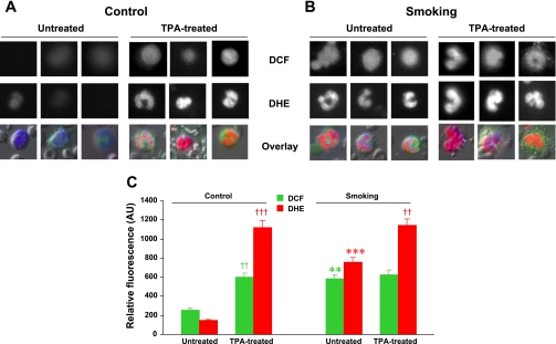
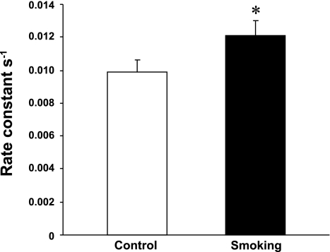
Cigarette smoking is a major independent risk factor for cardiovascular disease. While the association between chronic smoking and cardiovascular disease is well established, the underlying mechanisms are incompletely understood, partly due to the lack of adequate in vivo animal models. Here, we report a mouse model of chronic smoking-induced cardiovascular pathology. Male C57BL/6J mice were exposed to whole body mainstream cigarette smoke (CS) using a SCIREQ "InExpose" smoking system (48 min/day, 5 days/wk) for 16 or 32 wk. Age-matched, air-exposed mice served as nonsmoking controls. Blood pressure was measured, and cardiac MRI was performed. In vitro vascular ring and isolated heart experiments were performed to measure vascular reactivity and cardiac function. Blood from control and smoking mice was studied for the nitric oxide (NO) decay rate and reactive oxygen species (ROS) generation. With 32 wk of CS exposure, mice had significantly less body weight gain and markedly higher blood pressure. At 32 wk of CS exposure, ACh-induced vasorelaxation was significantly shifted to the right and downward, left ventricular mass was significantly larger along with an increased heart-to-body weight ratio, in vitro cardiac function tended to be impaired with high afterload, white blood cells had significantly higher ROS generation, and the blood NO decay rate was significantly faster. Thus, smoking led to blunted weight gain, hypertension, endothelial dysfunction, leukocyte activation with ROS generation, decreased NO bioavailability, and mild cardiac hypertrophy in mice that were not otherwise predisposed to disease. This mouse model is a useful tool to enable further elucidation of the molecular and cellular mechanisms of smoking-induced cardiovascular diseases.


Figures

Fig. 1.
Fig. 1.
Effects of cigarette smoke (CS) exposure on body weight (BW) and blood pressure (BP) under normal physiological conditions. Mice were divided into air-exposed (control) and CS-exposed (smoking) groups. CS exposure for 16 wk had no significant effects, but 32 wk of CS exposure significantly blunted BW gain (A) and increased BP (B) compared with control mice. Values are means ± SE; n = 5–7 mice/group. **P < 0.01 and ***P < 0.001 vs. control.
Fig. 2.
Fig. 2.
Effects of CS exposure on in vitro vascular endothelial function of the mouse aorta. CS exposure for 16 wk had no effects (A), but 32 wk of CS exposure significantly shifted ACh-induced aortic relaxation to the right and downward (B). PHE, phenylephrine. Values are means ± SE; n = 5 mice/group. *P < 0.05 vs. the respective control dose.
Fig. 3.
Fig. 3.
Effects of 32 wk of CS exposure on cardiac mass and contractile parameters. Left ventricular (LV) mass (A) and the heart weight-to-BW ratio (HW/BW; B) were significantly larger in CS-exposed mice compared with control mice. C: LV pressure-volume (P-V) relationship, which exhibited a trend toward LV systolic and diastolic dysfunction at high afterload in CS-exposed mice (S) compared with control mice (C). LVDP, LV developed pressure; LVEDP, LV end-diastolic pressure. Values are means ± SE; n = 5 mice/group. *P < 0.05 and **P < 0.01 vs. control.
Fig. 4.
Fig. 4.
Effects of 32 wk of CS exposure on ROS generation in mononuclear cells. A and B: representative mononuclear cells from control (A) and smoking (B) mice showing the fluorescence of 2′,7′-dichlorofluorescein (DCF) and dihydroethidium (DHE) as grayscale intensities at the top and middle rows, respectively. The bottom row shows the overlay with Hoechst (nuclei; blue), DCF (green), and DHE (red) fluorescences at the original magnification (×20). C: bar graphs of mean fluorescence intensities in untreated and 22-O-tetradecanoylporbol 13-acetate (TPA)-treated mononuclear cells from control and CS-exposed mice. In control cells, TPA induced activation with increased ROS generation (††P < 0.002, TPA-treated vs. respective untreated cells). For untreated cells, smoking induced clear activation with enhanced ROS generation (***P < 0.001 and **P < 0.002 vs. untreated cells, respectively, for DCF and DHE). For the smoking group, TPA induced only mild further increase in ROS, indicating that these cells are basally activated. AU, arbitrary units. Values are means ± SE; n = 8 samples/group.
Fig. 5.
Fig. 5.
Effects of 32 wk of CS exposure on ROS generation in polymorphonuclear cells. A and B: representative polymorphonuclear cells from control (A) and smoking (B) mice showing the fluorescence of DCF and DHE as grayscale intensities at the top and middle rows, respectively. The bottom row shows the overlay with Hoechst (nuclei; blue), DCF (green), and DHE (red) fluorescences at the original magnification (×20). C: bar graphs showing mean fluorescence intensities in untreated and TPA-treated polymorphonuclear cells. In control cells, TPA induced marked activation with increased ROS generation [††P < 0.002 (DCF) and †††P < 0.001 (DHE) for TPA-treated vs. respective untreated cells]. For untreated cells, smoking induced clear activation with enhanced ROS generation (**P < 0.002 and ***P < 0.001 vs. untreated cells, respectively, for DCF and DHE). For the smoking group, TPA induced further increases in ROS [††P < 0.002 (DHE)], indicating that these white blood cells are basally activated by CS exposure but still capable of further activation by TPA. Values are means ± SE; n = 8 samples/group.
Fig. 6.
Fig. 6.
Effects of 32 wk of CS exposure on the nitric oxide (NO) decay rate in red blood cells/white blood cells of control and CS-exposed mice. NO decay rate constants in diluted (1:3,000) blood samples from mice in the CS-exposed and control groups were determined by carbon fiber electrodes. Normalized calculated values show that the rate constant of NO decay in CS-exposed mice was greater compared with control mice. Values are means ± SE; n = 8 samples/group. *P < 0.05 vs. the respective control.

Similar articles

Cited by

References

    1. Ambrose JA, Barua RS. The pathophysiology of cigarette smoking and cardiovascular disease: an update. J Am Coll Cardiol 43: 1731–1737, 2004 - PubMed
    1. Barua RS, Ambrose JA, Eales-Reynolds LJ, DeVoe MC, Zervas JG, Saha DC. Dysfunctional endothelial nitric oxide biosynthesis in healthy smokers with impaired endothelium-dependent vasodilatation. Circulation 104: 1905–1910, 2001 - PubMed
    1. Bergmann S, Siekmeier R, Mix C, Jaross W. Even moderate cigarette smoking influences the pattern of circulating monocytes and the concentration of sICAM-1. Respir Physiol 114: 269–275, 1998 - PubMed
    1. Bernhard D, Wang XL. Smoking, oxidative stress and cardiovascular diseases–do anti-oxidative therapies fail? Curr Med Chem 14: 1703–1712, 2007 - PubMed
    1. Brook RD, Rajagopalan S. Particulate matter, air pollution, and blood pressure. J Am Soc Hypertens 3: 332–350, 2009 - PubMed

Publication types

MeSH terms