. 2010 Nov 23;107(47):20269-74.
doi: 10.1073/pnas.1004783107. Epub 2010 Nov 5.

Archaeal-like chaperonins in bacteria

Affiliations

Archaeal-like chaperonins in bacteria

Stephen M Techtmann et al. Proc Natl Acad Sci U S A. .

Abstract

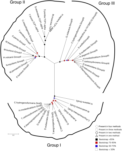
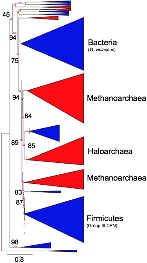
Chaperonins (CPN) are ubiquitous oligomeric protein machines that mediate the ATP-dependent folding of polypeptide chains. These chaperones have not only been assigned stress response and normal housekeeping functions but also have a role in certain human disease states. A longstanding convention divides CPNs into two groups that share many conserved sequence motifs but differ in both structure and distribution. Group I complexes are the well known GroEL/ES heat-shock proteins in bacteria, that also occur in some species of mesophilic archaea and in the endosymbiotic organelles of eukaryotes. Group II CPNs are found only in the cytosol of archaea and eukaryotes. Here we report a third, divergent group of CPNs found in several species of bacteria. We propose to name these Group III CPNs because of their distant relatedness to both Group I and II CPNs as well as their unique genomic context, within the hsp70 operon. The prototype Group III CPN, Carboxydothermus hydrogenoformans chaperonin (Ch-CPN), is able to refold denatured proteins in an ATP-dependent manner and is structurally similar to the Group II CPNs, forming a 16-mer with each subunit contributing to a flexible lid domain. The Group III CPN represent a divergent group of bacterial CPNs distinct from the GroEL/ES CPN found in all bacteria. The Group III lineage may represent an ancient horizontal gene transfer from an archaeon into an early Firmicute lineage. An analysis of their functional and structural characteristics may provide important insights into the early history of this ubiquitous family of proteins.


Conflict of interest statement

The authors declare no conflict of interest.

Figures

Fig. 1.
Fig. 1.
Phylogenetic analysis of Group III chaperonin: Unrooted minimum evolution phylogenetic tree representing Group I, Group II, and the new Group III chaperonins, indicating the great phylogenetic distance between the Group III clade and the conserved Group I and Group II clusters. The same alignment was used to construct trees using multiple algorithms (minimum evolution, maximum parsimony, and neighbor-joining). Nodes present in trees created by different alogorithms and with high bootstrap values are denoted as described. The clade representing Group III CPN contains the following organisms: Carboxydothermus hydrogenoformans, Geobacillus spY412MC, Syntrophomonas. wolfei wolfei Goettingen, Desulforudis audaxviator, Ammonifex degensii, Heliobacterium modesticaldum, Thermosinus carboxydivorans, Desulfotomaculum acetoxidans, Desulfotomaculum reducens, Pelotomaculum thermopropionicum, and Gloeobacter violaceus.
Fig. 2.
Fig. 2.
Genomic context of the Group III chaperonin: (A) The genomic context of all but one of the Group III chaperonins. As shown, the Group III CPN (cpn) is downstream of hrcA and is adjacent to the Hsp70 operon (dnaK, dnaJ, grpE) This topology is found in C. hydrogenoformans, Geobacillus spY412MC, S. wolfei wolfei Goettingen, D. audaxviator, A. degensii, H. modesticaldum, T. carboxydivorans, D. acetoxidans, D. reducens, and P. thermopropionicum. (B) The genomic context of G. violaceus. This CPN is not in close association with any other chaperones. (C) The CIRCE sequences upstream of the Group I and Group III CPN from C. hydrogenoformans.
Fig. 3.
Fig. 3.
Chaperone activity: (A) RT-PCR of the Group I and Group III chaperonins from C. hydrogenoformans. (B) GDH protection assays with Ch-CPN at both 42 °C (closed symbols) and 50 °C (open symbols). One No-ATP control was performed at 50 °C (open triangles). (C) Malate dehydrogenase refolding assays. Denatured MDH protein was mixed with Ch-CPN and ATP to determine Ch-CPN’s ability to refold denatured proteins. The original (predenaturation) activity was used to determine 100%. No MDH refolding was observed with Ch-CPN alone (open symbols). Refolding was observed when 0.5 M AS was added (closed symbols). (D) E. coli survival assays. E. coli BL21 expressing Ch-CPN (open symbols) and E. coli BL21 with empty vector (closed symbols) were heated to 50 °C for 2 h. Viable counts were taken at time intervals.
Fig. 4.
Fig. 4.
Evidence for a helical protrusion in the Group III chaperonin. (A) Structural model of C. hydrogenoformans CPN based on the structure of the T. acidophilum Group II chaperonin, constructed using the Wurst program. The structure is colored based on conservation to all CPN in the alignment. Molecular graphics images were produced using the UCSF Chimera package from the Resource for Biocomputing, Visualization, and Informatics at the University of California, San Francisco (49). (B) Native PAGE of Ch-CPN incubated at 65 °C with various components of buffer A and ATP. The two bands representing two forms of Ch-CPN (open and closed). Upon addition of ATP only the open form is observed. (C) Representative class averaged images of side-views of Ch-CPN elucidating the presence of built-in lid (indicated by white arrows). These side views are averages of 411, 353, and 298 images respectively. Top view is the average of 245 images. All of the classes generated during image averaging are in (Fig. S5).
Fig. 5.
Fig. 5.
Phylogenetic tree of DnaK homologs from Group III CPN-containing organisms. A maximum liklihood tree was constructed using the PhyML program. DnaK sequences from the Group III CPN gene cluster were aligned with all archaeal DnaK homologs and those of some bacteria. Clades composed of all bacteria are colored blued. Clades comprised of archaea are colored red. The Group III CPN-associated DnaKs cluster along with other Firmicutes in a clade distinct from any archaeal DnaKs. (Full tree is in Fig. S6)

References

    1. Laksanalamai P, Robb FT. Small heat shock proteins from extremophiles: a review. Extremophiles. 2004;8:1–11. - PubMed
    1. Laksanalamai P, Whitehead TA, Robb FT. Minimal protein-folding systems in hyperthermophilic archaea. Nat Rev Microbiol. 2004;2:315–324. - PubMed
    1. Large AT, Goldberg MD, Lund PA. Chaperones and protein folding in the archaea. Biochem Soc Trans. 2009;37:46–51. - PubMed
    1. Large AT, Lund PA. Archaeal chaperonins. Front Biosci. 2009;14:1304–1324. - PubMed
    1. Macario AJ, Conway De Macario E. The molecular chaperone system and other anti-stress mechanisms in archaea. Front Biosci. 2001;6:D262–283. - PubMed

Publication types

MeSH terms

LinkOut - more resources