. 2010 Dec;7(12):995-1001.
doi: 10.1038/nmeth.1529. Epub 2010 Nov 7.

FragSeq: transcriptome-wide RNA structure probing using high-throughput sequencing

Affiliations

FragSeq: transcriptome-wide RNA structure probing using high-throughput sequencing

Jason G Underwood et al. Nat Methods. 2010 Dec.

Abstract

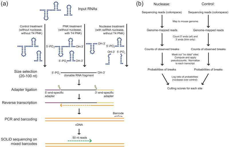
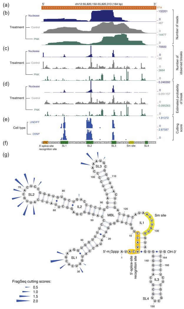
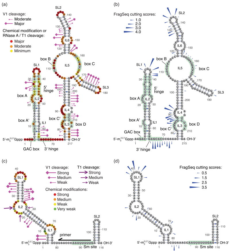
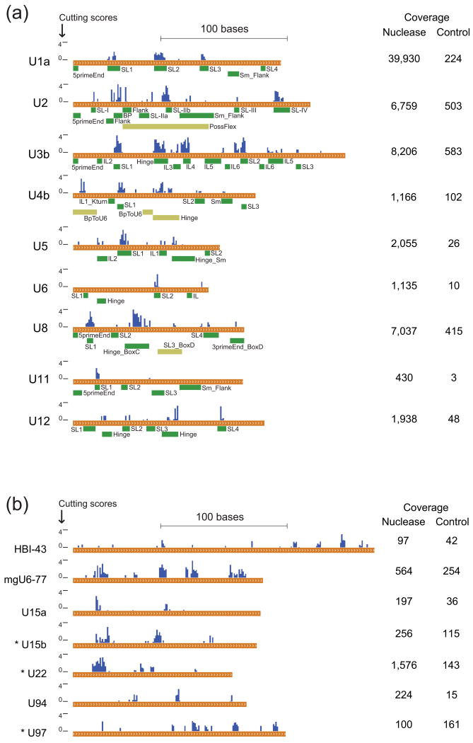
Classical approaches to determine structures of noncoding RNA (ncRNA) probed only one RNA at a time with enzymes and chemicals, using gel electrophoresis to identify reactive positions. To accelerate RNA structure inference, we developed fragmentation sequencing (FragSeq), a high-throughput RNA structure probing method that uses high-throughput RNA sequencing of fragments generated by digestion with nuclease P1, which specifically cleaves single-stranded nucleic acids. In experiments probing the entire mouse nuclear transcriptome, we accurately and simultaneously mapped single-stranded RNA regions in multiple ncRNAs with known structure. We probed in two cell types to verify reproducibility. We also identified and experimentally validated structured regions in ncRNAs with, to our knowledge, no previously reported probing data.


Figures

Figure 1
Figure 1. Overview of the FragSeq method
a, Preparation of FragSeq libraries for sequencing. RNA 5′ and 3′ end chemistry is specifically shown to highlight PNK and nuclease products; when RNA end chemistry is not shown, it denotes any possible end chemistry. Only clonable RNA fragments are shown at and after the size-selection step. Lightning bolts represent the specific ligation events at each end of the RNA fragment. b, Overview of the FragSeq algorithm.
Figure 2
Figure 2. Visual representation of data at progressive stages in the FragSeq algorithm, from genome-mapped reads to cutting scores
a-e, Data tracks in the UCSC Genome Browser (mm9 mouse genome assembly) showing spliceosomal snRNA U1a (a); data from mouse undifferentiated embryonic stem cell samples (UNDIFF) (b-d) is processed to get cutting scores, which are compared to cutting scores from D5NP cells (e). Ignored sites (Supplementary Note 1) are denoted in (e) as areas for which no data is shown (e.g. the sequence GUG in the Sm region). f, Sequence of U1a, highlighting regions shown in (g) using the same color code; green and yellow subsequences are expected to be single-stranded. g, Cutting scores (blue arrows) from UNDIFF sample (e) superimposed on the known secondary structure. Non-canonical base pairs in interior loops of stem 2 are shown as unpaired. 2′-O-methylated positions are not depicted. SL, stem-loop; IL, interior loop; MBL, multibranch loop. U1a structure is from several sources (Supplementary Note 3).
Figure 3
Figure 3. Comparison of FragSeq with previous probing experiments
a-b, Probing results for human U3 purified from HeLa cells (a) and FragSeq cutting scores for mouse U3b (b). c-d, Probing results for human U5 purified from HeLa cells (c) and FragSeq cutting scores for mouse U5 (d). Black arrow shows priming position for primer extension; only bases downstream of the primer were probed in that study (c). Reactivities in (a) and (c) are taken verbatim from ref. 15 and ref. 16, respectively; structures and other annotations were compiled from multiple sources (Supplementary Note 3). 2′-O-methylated positions are not depicted.
Figure 4
Figure 4. FragSeq cutting scores and coverage for ncRNAs with known structures and long C/D box snoRNAs
Coverage (mean reads per nucleotide) is shown at right for nuclease and control treatments. a, Cutting scores compared to ssRNA regions greater than three bases long (green boxes) for ncRNAs with published structure models (Supplementary Note 3). Regions exist where the in vitro structure of a single, naked RNA is uncertain (olive boxes). SL, stem-loop; Sm, Sm protein binding site; BP, splicing branch-point binding site; Flank, flanking ssRNA region of a nearby motif; IL, interior loop; Hinge, ssRNA region connecting two RNA domains; Kturn, kink-turn RNA motif containing non-canonical base pairs. b, Cutting scores for all long (> 120nt) C/D box snoRNAs considered for follow-up probing. RNAs with an asterisk (*) were chosen for follow-up probing.
Figure 5
Figure 5. FragSeq probing versus conventional nuclease probing of U15b C/D box snoRNA
a, FragSeq ssRNA cutting scores (bottom, dark blue) and band quantification readouts (SAFA counts) based on the gel resolving 5′-end-labeled probing products. X-axis shows nucleotide numbering; gridlines appear every five nucleotides. Gray nucleotides in sequence show areas that were outside of the reliably quantifiable area on the gel. Parentheses denote Watson/Crick base pairs and dots denote ssRNA. Triangles denote bases where a nuclease can cut: T1, gray triangles at G; RNase A, black triangles for C and red triangles for U. Outlier values were truncated and marked with red zigzag lines. b, Follow-up probing data superimposed on our structure model, with probing enzymes color-coded as in (a). Marginal, weak, moderate, or strong enzyme activity was inferred from manual inspection of the gel and Safa quantification from (a) (Supplementary Note 3). c, FragSeq cutting scores superimposed on the same structure model as (a) and (b). Boxes (green) and the putative region that base-pairs with target rRNA (orange) are highlighted, with the base opposite of the methylated position in red. Highlighting is as in (b).

References

    1. Gesteland R, Cech T, Atkins J, editors. The RNA World. 3rd. Cold Spring Harbor Laboratory Press; Cold Spring Harbor, New York: 2005.
    1. Affymetrix/Cold Spring Harbor Laboratory ENCODE Transcriptome Project. Post-transcriptional processing generates a diversity of 5′-modified long and short RNAs. Nature. 2009;457:1028–1032. - PMC - PubMed
    1. Guttman M, et al. Chromatin signature reveals over a thousand highly conserved large non-coding RNAs in mammals. Nature. 2009;458:223–227. - PMC - PubMed
    1. Ambros V. microRNAs: tiny regulators with great potential. Cell. 2001;107:823–826. - PubMed
    1. Kapranov P, et al. RNA maps reveal new RNA classes and a possible function for pervasive transcription. Science. 2007;316:1484–1488. - PubMed

Publication types