. 2010 Dec;11(12):1136-42.
doi: 10.1038/ni.1960. Epub 2010 Nov 7.

Caspase-1-induced pyroptosis is an innate immune effector mechanism against intracellular bacteria

Affiliations

Caspase-1-induced pyroptosis is an innate immune effector mechanism against intracellular bacteria

Edward A Miao et al. Nat Immunol. 2010 Dec.

Abstract

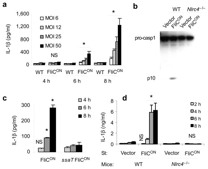
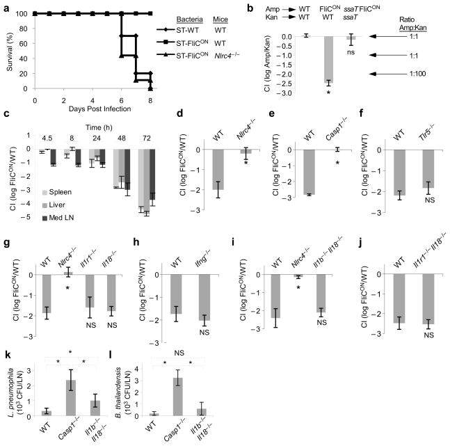
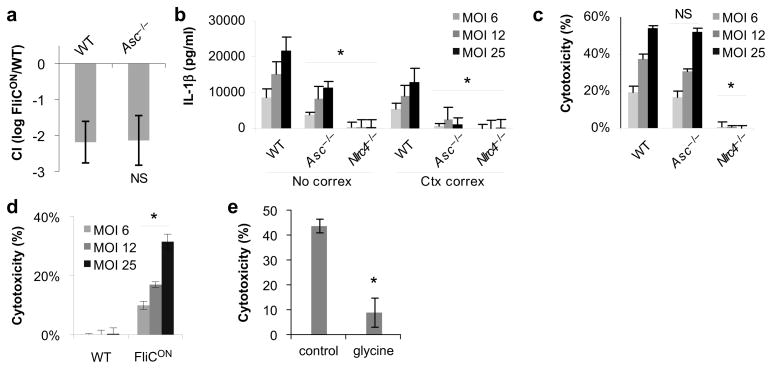
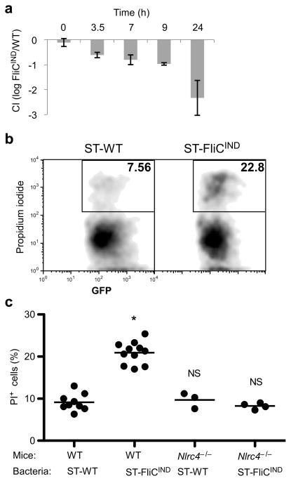
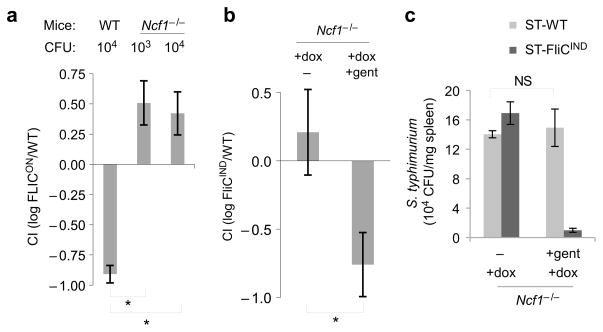
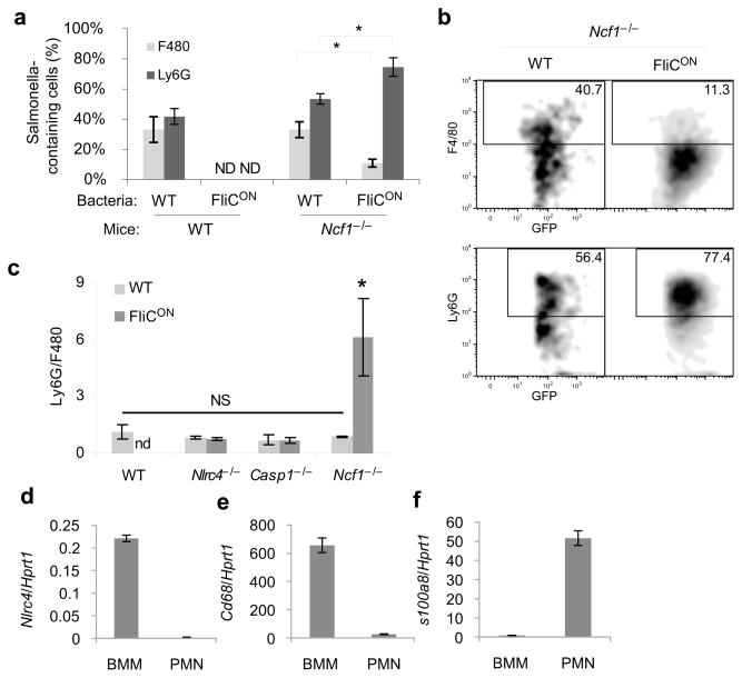
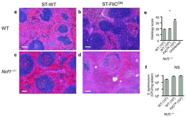
Macrophages mediate crucial innate immune responses via caspase-1-dependent processing and secretion of interleukin 1β (IL-1β) and IL-18. Although infection with wild-type Salmonella typhimurium is lethal to mice, we show here that a strain that persistently expresses flagellin was cleared by the cytosolic flagellin-detection pathway through the activation of caspase-1 by the NLRC4 inflammasome; however, this clearance was independent of IL-1β and IL-18. Instead, caspase-1-induced pyroptotic cell death released bacteria from macrophages and exposed the bacteria to uptake and killing by reactive oxygen species in neutrophils. Similarly, activation of caspase-1 cleared unmanipulated Legionella pneumophila and Burkholderia thailandensis by cytokine-independent mechanisms. This demonstrates that activation of caspase-1 clears intracellular bacteria in vivo independently of IL-1β and IL-18 and establishes pyroptosis as an efficient mechanism of bacterial clearance by the innate immune system.


Figures

Figure 1
Figure 1. Characterization of flagellin-expressing S. typhimurium
(a–d) BMDM were infected with ST-WT or ST-FliCON under conditions where SPI2 T3SS is expressed and SPI1 T3SS is not expressed at the indicated multiplicity of infection (MOI) for one hour followed by treatment with gentamicin. Total infection time is indicated. (a) IL-1β concentration determined by ELISA. (b) Caspase-1 processing analyzed by immunobloting. (c) IL-1β concentration after infection with ST-FliCON or SPI2 mutant ST-FliCON (ssaT) at MOI 12. (d) IL-1β concentration after infection of WT or Nlrc4−/−BMDM at MOI 10. Data is representative of at least three experiments. * = p < 0.05, NS = p > 0.05 from relevant control.
Figure 2
Figure 2. Flagellin expression attenuates S. typhimurium in vivo
(a) WT or Nlrc4−/−mice were infected with 100 colony forming units (CFU) ST-WT or ST-FliCON and survival was monitored. (b) WT mice were infected with equal numbers of two bacterial strains marked with different antibiotic markers as indicated. CFU in the spleen were determined after 48h and the log of the ratio is shown as the competitive index. Note that a competitive index of -2 corresponds to a 100-fold reduction in the ampicillin resistant strain. (c) Competitive index of ST-FliCON/ST-WT in various tissues. (d–j) Competitive index of ST-FliCON/ST-WT in the indicated WT or knock out mice at 48 h post infection. Samples marked with * are statistically significant from the other samples in the same graph (p < 0.05); ns: not significantly different from WT or other samples marked with “ns” (p > 0.05). (k, l) WT, Casp1−/−, or Il1b−/−Il18−/−mice were infected intraperitoneally with (k) L. pneumophila (data shown from n=4, 6, and 6 mice, respectively, representative of two experiments) or (l) B. thailandensis (data shown from n=6, 4, and 6 mice, respectively, representative of four experiments). CFU were enumerated 24 h later from the draining lymph nodes. * = p < 0.05, NS = p > 0.05. See Supplementary Table 1 for mouse numbers in panels b–j.
Figure 3
Figure 3. Differential role of ASC in NLRC4 signalling
(a) Competitive index of ST-FliCON vs. ST-WT as in Figure 2 for WT (n=6) and Asc−/−(n=4) mice. (b, c) BMDM from WT, Asc−/− or Nlrc4−/−mice were infected as in Figure 1 with ST-FliCON and IL-1β secretion or cytotoxicity were determined by ELISA and LDH release 6 hours post infection. (d) WT BMDM infected with ST-WT or ST-FliCON and pyroptosis measured by LDH release 8h post infection. (e) Cytotoxicity induced by ST-FliCON was monitored with or without 10 mM glycine added to the media and cytotoxicity determined 7h post infection. In vitro infections are representative of at least three experiments. * = p < 0.05, NS = p > 0.05 from relevant control.
Figure 4
Figure 4. Evidence for pyroptosis in vivo
(a–c) Mice were infected with either ST-WT (flgB GFP) or flagellin-inducible ST-FliCIND (flgB GFP pEM87) for 48 h, after which doxycycline was injected to induce flagellin expression. (a) Competitive index of ST-FliCIND/ST-WT after synchronized induction of flagellin expression (n=2, except 0h n=3). (b–c) GFP-containing cells from the peritoneal wash were analyzed by flow cytometry for membrane integrity by propidium iodide staining (PI). (b) Representative flow cytometry analysis and (c) percent PI positive bacteria containing cells from WT (n=9 and n=11 for ST-WT and ST-FliCIND) or Nlrc4−/− (n=3 and n=4 for ST-WT and ST-FliCIND) mice per group, average is shown (bar). * = p < 0.001, NS = p > 0.05.
Figure 5
Figure 5. ST-FliCON are released from macrophages and cleared by ROS
(a) Competitive index of ST-FliCON/ST-WT 24 h post infection in WT or Ncf1−/−mice infected with the indicated inoculum (n=2, except n=3 for Ncf1−/−103 CFU). (b) Competitive index and (c) individual CFU from coinfection of Ncf1−/−mice with ST-FliCIND/ST-WT on a flgB mutant background (which ablate endogenous flagellin expression which occurs during growth in LB broth); 24h post infection mice were injected with doxycycline alone (n=8) or doxycycline plus gentamicin (n=10), and CFU were determined 24h later. * = p < 0.05, NS = p > 0.05.
Figure 6
Figure 6. ST-FliCON accumulate within neutrophils
(a–c) The indicated mouse strains were infected with GFP-expressing ST-WT or ST-FliCON for 48 hours (n=4 for each; results are representative of three experiments). GFP-containing splenocytes were interrogated for macrophage (F4/80) or neutrophils (Ly6G) markers by flow cytometry. (a) Percent marker positive cells and (b) representative individual plot are shown. (c) Ratio of GFP+ Ly6G+ to GFP+ F4/80+ cells determined by flow cytometry. (d–f) qPCR from BMDM or neutrophils (PMNs) for NLRC4, or macrophage (CD68) or neutrophil (S100a8) specific marker controls representative of three experiments. * = p < 0.05, NS = p > 0.05, ND = not detectable.
Figure 7
Figure 7. Sustained NLRC4 activation causes tissue damage
WT or Ncf1−/−mice were infected with ST-WT or ST-FliCON for 48 h. WT mice were infected with 105 CFU of each strain. Ncf−/− mice infected with 103 or 104 CFU ST-WT, or 103 CFU ST-FliCON; note that bacterial loads 48h post infection were similar between mice infected with 104 CFU ST-WT and 103 CFU ST-FliCON (see f). (a–d) Histopathology of spleens from WT or Ncf1−/−mice infected with ST-WT or ST-FliCON. WT mice infected wit ST-WT (a) had multifocal neutrophilic to necrosuppurative splenitis and red pulp congestion. In contrast, WT mice infected with ST-FliCON (b) had nearly normal splenic morphology. Splenic lesions in the Ncf1−/−mice infected with 103 ST-FliCON (d) were severe and consisted of marked red and white pulp necrosis. Less severe changes were seen in Ncf1−/−mice infected with 104 ST-WT (c), including red pulp congestion, thrombosis, neutrophilic to necrosuppurative splenitis, while the white pulp was relatively spared. Scale bar = 100μm. (e) Scoring of pathologic changes in spleens from Ncf1−/−mice (n=3 per inoculum). (f) CFU from the spleen were determined (n=3 per inoculum). Two doses of ST-WT were used in Ncf1−/−mice in order to compare to ST-FliCON infected mice normalized for either initial dose or 48h bacterial load; Ncf1−/−infected with 104 CFU ST-WT have similar bacterial loads to Ncf1−/−mice infected with 103 ST-FliCON. * = p < 0.05, NS = p > 0.05.

Similar articles

Cited by

References

    1. Miao EA, Andersen-Nissen E, Warren SE, Aderem A. TLR5 and Ipaf: dual sensors of bacterial flagellin in the innate immune system. Semin Immunopathol. 2007;29:275–288. - PubMed
    1. Dinarello CA. Immunological and inflammatory functions of the interleukin-1 family. Annu Rev Immunol. 2009;27:519–550. - PubMed
    1. Szretter KJ, et al. Role of host cytokine responses in the pathogenesis of avian H5N1 influenza viruses in mice. J Virol. 2007;81:2736–2744. - PMC - PubMed
    1. Schmitz N, Kurrer M, Bachmann MF, Kopf M. Interleukin-1 is responsible for acute lung immunopathology but increases survival of respiratory influenza virus infection. J Virol. 2005;79:6441–6448. - PMC - PubMed
    1. Kozak W, et al. Thermal and behavioral effects of lipopolysaccharide and influenza in interleukin-1 beta-deficient mice. Am J Physiol. 1995;269:R969–977. - PubMed

Publication types

MeSH terms