. 2011 Mar 10;30(10):1159-73.
doi: 10.1038/onc.2010.497. Epub 2010 Nov 8.

Absence of the Birt-Hogg-Dubé gene product is associated with increased hypoxia-inducible factor transcriptional activity and a loss of metabolic flexibility

Affiliations

Absence of the Birt-Hogg-Dubé gene product is associated with increased hypoxia-inducible factor transcriptional activity and a loss of metabolic flexibility

R S Preston et al. Oncogene. .

Abstract

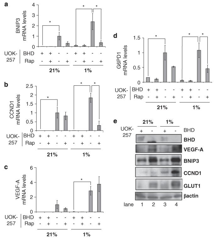
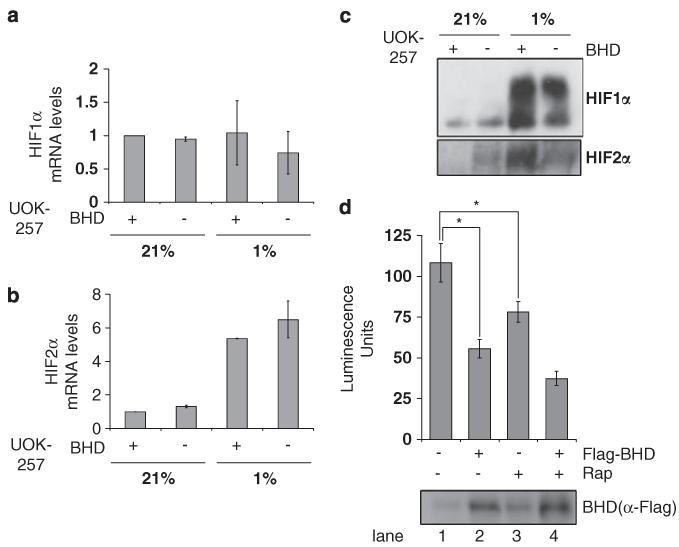
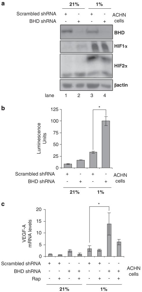
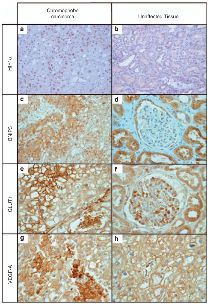
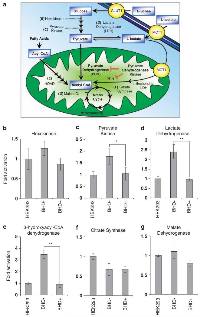
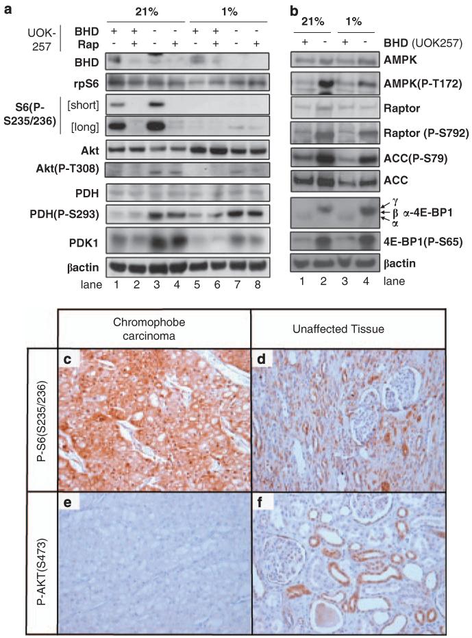
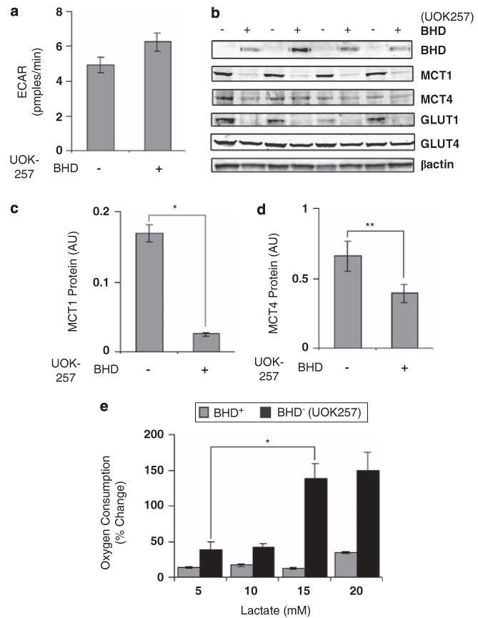
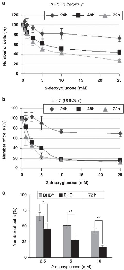
Under conditions of reduced tissue oxygenation, hypoxia-inducible factor (HIF) controls many processes, including angiogenesis and cellular metabolism, and also influences cell proliferation and survival decisions. HIF is centrally involved in tumour growth in inherited diseases that give rise to renal cell carcinoma (RCC), such as Von Hippel-Lindau syndrome and tuberous sclerosis complex. In this study, we examined whether HIF is involved in tumour formation of RCC in Birt-Hogg-Dubé syndrome. For this, we analysed a Birt-Hogg-Dubé patient-derived renal tumour cell line (UOK257) that is devoid of the Birt-Hogg-Dubé protein (BHD) and observed high levels of HIF activity. Knockdown of BHD expression also caused a threefold activation of HIF, which was not as a consequence of more HIF1α or HIF2α protein. Transcription of HIF target genes VEGF, BNIP3 and CCND1 was also increased. We found nuclear localization of HIF1α and increased expression of VEGF, BNIP3 and GLUT1 in a chromophobe carcinoma from a Birt-Hogg-Dubé patient. Our data also reveal that UOK257 cells have high lactate dehydrogenase, pyruvate kinase and 3-hydroxyacyl-CoA dehydrogenase activity. We observed increased expression of pyruvate dehydrogenase kinase 1 (a HIF gene target), which in turn leads to increased phosphorylation and inhibition of pyruvate dehydrogenase. Together with increased protein levels of GLUT1, our data reveal that UOK257 cells favour glycolytic rather than lipid metabolism (a cancer phenomenon termed the 'Warburg effect'). UOK257 cells also possessed a higher expression level of the L-lactate influx monocarboxylate transporter 1 and consequently utilized L-lactate as a metabolic fuel. As a result of their higher dependency on glycolysis, we were able to selectively inhibit the growth of these UOK257 cells by treatment with 2-deoxyglucose. This work suggests that targeting glycolytic metabolism may be used therapeutically to treat Birt-Hogg-Dubé-associated renal lesions.


Figures

Figure 1
Figure 1
BHD negatively regulates HIF-induced gene expression during hypoxia. The mRNA levels of (a) BNIP3, (b) CCND1, (c) VEGF-A and (d) G6PD1 were compared in BHD+ (UOK257-2) and BHD (UOK257) cells treated overnight with 50 nM rapamycin under normoxia (21%) and hypoxia (1%), where indicated, in this study by quantitative reverse-transcriptase PCR. mRNA levels were standardized against β-Actin and fold activation was compared with that in BHD cells under normoxia. n = 3. *P<0.05 when comparing BHD+ and BHD cells under hypoxia and normoxia and rapamycin treatment. (e) Western blot analysis was carried out on cell lysates prepared from BHD+ and BHD cells under normoxia (21%) and hypoxia (1%), where indicated. Protein levels of BHD, VEGF-A, BNIP3, CCND1, GLUT1 and β-actin (as loading control) were determined.
Figure 2
Figure 2
Increased transcriptional activity of HIF1α and HIF2α in BHD cells. The mRNA of (a) HIF1α and (b) HIF2α was compared in BHD+ (UOK257-2) and BHD (UOK257) cells after 18 h of normoxia (21%) or hypoxia (1%), where indicated, by reverse transcriptase PCR. mRNA levels were standardized against β-actin and fold activation was compared with that in BHD+ cells under normoxia. n = 3. (c) The protein levels of both HIF1α and HIF2α were analysed by western blot analysis (d) BHD cells transiently transfected with an HIF-inducible luciferase reporter and either empty pRK7 or Flag-BHD vector and then treated with 50nm rapamycin ‘Rap’ (where indicated) were maintained at 1% O2 for 18 h. Lysates were analysed for luciferase fluorescence to determine the transcriptional activity of HIF. n = 3. *P<0.05. Protein levels of Flag-tagged BHD were determined with anti-Flag antibodies.
Figure 3
Figure 3
Knockdown of BHD expression increases HIF activity under hypoxia in human kidney ACHN cells. (a) The protein levels of HIF1α and HIF2α were compared in ACHN cells, stably transfected with either BHD shRNA or scrambled shRNA, after 18 h of normoxia (21%) or hypoxia (1%) by western blot. β-actin serves as a loading control and endogenous BHD expression was determined to verify efficient BHD knockdown. (b) ACHN cells, stably transfected with either BHD shRNA or scrambled shRNA were transiently transfected with an HIF-inducible luciferase reporter and then maintained at either 21 or 1% O2 for 18 h. Lysates were analysed for luciferase fluorescence to determine the transcriptional activity of HIF. n = 3. *P<0.05. (c) The mRNA levels of VEGF-A were determined from ACHN/scrambled-shRNA and ACHN/BHD-shRNA cells that were treated overnight with 50 nm rapamycin under normoxia (21%) and hypoxia (1%), where indicated, by quantitative reverse-transcriptase PCR. mRNA levels were standardized against β-actin. Fold activation was compared with that in ACHN/BHD-shRNA cells under normoxia. n = 3. *P<0.05 when comparing ACHN cells lines under hypoxia.
Figure 4
Figure 4
Increased levels of HIF target proteins. Paraffin-embedded samples were obtained from a chromophobe renal carcinoma from a patient with Birt–Hogg–Dubé. There is strong and specific staining with antibodies directed against HIF1α (a, b) and the HIF targets BNIP3 (c, d), GLUT1 (e, f) and VEGF-A (g, h). Magnification is ×400 for all panels, apart from the HIF1α panels that are at ×200. Note the different staining patterns for BNIP3 and GLUT1 in chromophobe carcinoma compared with unaffected tissue. For VEGF, staining is much more intense in the tumour than in normal tissue but the intracellular distribution seems to be more diffuse. (e, f) Erythrocytes are clearly stained, serving as a positive internal control. In panels (g) and (h) vascular endothelium does the same for VEGF.
Figure 5
Figure 5
Increased enzyme activity levels of pyruvate kinase and lactate dehydrogenase in BHD cells. (a) Diagram of glucose and fatty acid metabolism depicting aerobic and anaerobic respiration. Enzyme activity assays were conducted to compare activity levels of (b) hexokinase, (c) pyruvate kinase, (d) lactate dehydrogenase, (e) 3-hydroxyacyl-CoA dehydrogenase, (f) citrate synthase, (g) malate dehydrogenase in BHD (UOK257), BHD+ (UOK257-2) and HEK-293 cells. n = 3. *P<0.01; **P<0.05 when comparing enzyme activity in BHD+ and BHD cells.
Figure 6
Figure 6
Inactivation of PDH and activation of mTOR and AMPK in BHD cells. (a) Western blot analyses were carried out on cell lysates obtained from BHD+ (UOK257-2) and BHD (UOK257) cells after 18 h of normoxia (21%) or hypoxia (1%), with and without 50 nm rapamycin, where indicated. Protein levels of BHD, rpS6, phosphorylated rpS6 at Ser235 and Ser236, Akt, phosphorylated Akt at Thr308, PDH, phosphorylated PDH at Ser293, PDK1 and β-actin (used as a loading control) were determined. (b) Western blot analyses were carried out on cell lysates obtained from BHD+ (UOK257-2) and BHD (UOK257) cells after 18 h of normoxia (21%) or hypoxia (1%), where indicated. Protein levels of AMPKα, phosphorylated AMPKα at Thr172, raptor, phosphorylated raptor at Ser792, acetyl-CoA carboxylase (ACC), phosphorylated ACC at Ser79, 4E-BP1, phosphorylated 4E-BP1 at Ser65 and β-actin (used as a loading control) were determined. Chromophobe renal carcinoma paraffin-embedded samples from a Birt–Hogg–Dubé patient show a strong and specific staining with phosphorylated rpS6 (c, d) but not Akt (e, f) antibodies. Magnification is ×200 for all panels.
Figure 7
Figure 7
BHD cells utilize l-lactate as a metabolic fuel. (a) The extracellular acidification rate (ECAR) was determined in BHD+ (UOK257-2) and BHD (UOK257) cells under normoxia. (b) Western blot analyses were performed to determine protein levels of BHD, MCT1, MCT4, GLUT1 and GLUT4 from lysates prepared from BHD and BHD+ grown in normoxia. Lysates from four different experiments were analysed. β-actin was used as a loading control. Densitometry analyses were also performed on (c) MCT1 and (d) MCT4 for the purpose of comparison. n = 4. *P<0.05; **P<0.01 relative to BHD+ and BHD cells. (e) Oxygen consumption was also measured in BHD+ and BHD cells treated with 5, 10, 15 and 20 mm l-lactate. n = 3. *P<0.05 relative to oxygen consumption within BHD cells at 5 mm and 15 mm l-lactate.
Figure 8
Figure 8
Proliferation of BHD cells is selectively inhibited with 2-deoxyglucose. (a) BHD+ (UOK257-2) and (b) BHD (UOK257) were treated with 0, 1.25, 2.5, 5, 10 and 25 mm 2-deoxyglucose. The percentage of the original number of cells present in each cell line was then determined after 24, 48 and 72 h. n = 3. (c)The percentage of the number of BHD+ and BHD cells after 72 h of treatment with 2.5, 5 and 10 mm 2-deoxyglucose from a and b was directly compared. n = 3. *P<0.05; **P<0.01.

References

    1. Baba M, Hong S-B, Sharma N, Warren MB, Nickerson ML, Iwamatsu A, et al. Folliculin encoded by the BHD gene interacts with a binding protein, FNIP1, and AMPK, and is involved in AMPK and mTOR signalling. P Natl Acad Sci USA. 2006;103:15552–15557. - PMC - PubMed
    1. Bratslavsky G, Sudarshan S, Neckers L, Linehan WM. Pseudohypoxic pathways in renal cell carcinoma. Clin Cancer Res. 2007;13:4667–4671. - PubMed
    1. Bustin SA, Benes V, Garson JA, Hellemans J, Huggett J, Kubista M, et al. The MIQE guidelines: minimum information for publication of quantitative real-time PCR experiments. Clin Chem. 2009;55:611–622. - PubMed
    1. Chen Z, Lu W, Garcia-Prieto C, Huang P. The Warburg effect and its cancer therapeutic implications. J Bioenerg Biomembr. 2007;39:267–274. - PubMed
    1. Deldicque L, Theisen D, Bertrand L, Hespel P, Hue L, Francaux M. Creatine enhances differentiation of myogenic C2C12 cells by activating both p38 and Akt/PKB pathways. Am J Physiol. 2007;293:1263–1271. - PubMed

Publication types

MeSH terms

Substances