Comparative Study
. 2011 Mar 17;30(11):1360-71.
doi: 10.1038/onc.2010.506. Epub 2010 Nov 8.

p85α mediates p53 K370 acetylation by p300 and regulates its promoter-specific transactivity in the cellular UVB response

Affiliations
Comparative Study

p85α mediates p53 K370 acetylation by p300 and regulates its promoter-specific transactivity in the cellular UVB response

L Song et al. Oncogene. .

Abstract

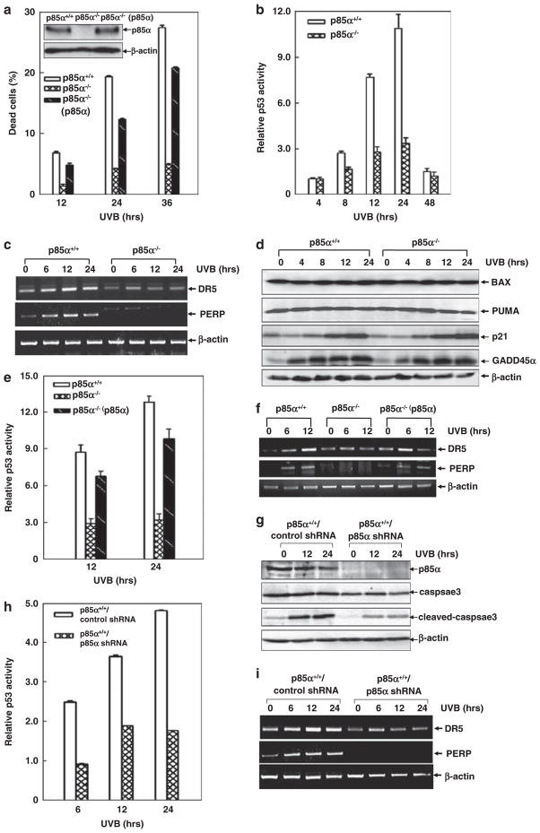
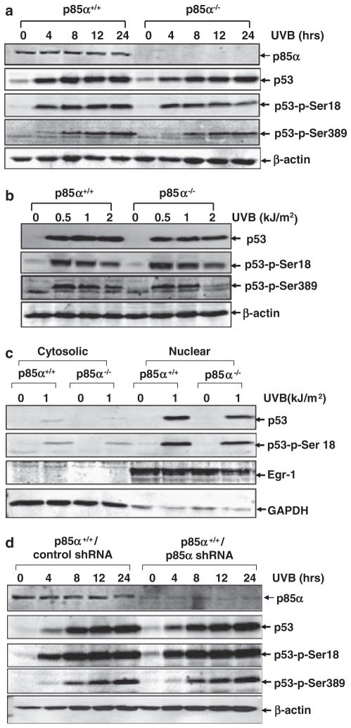
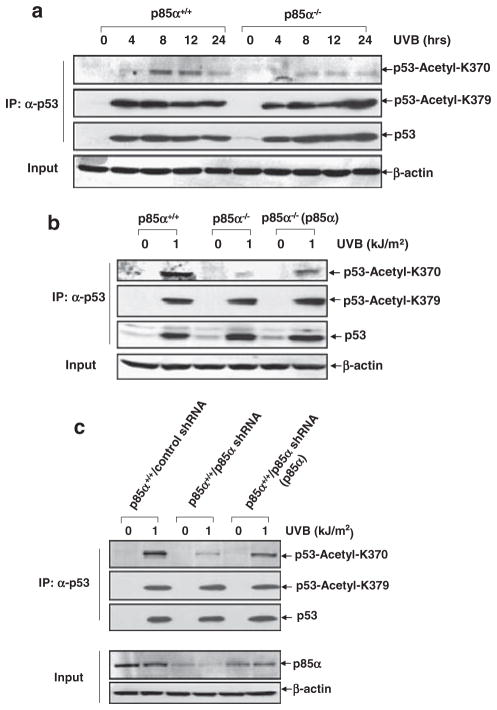
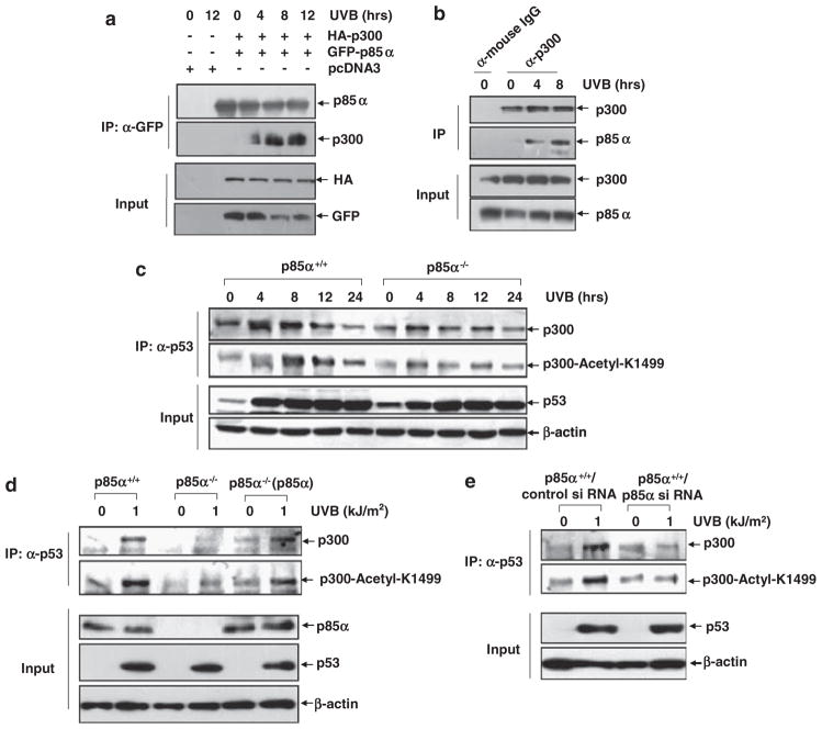
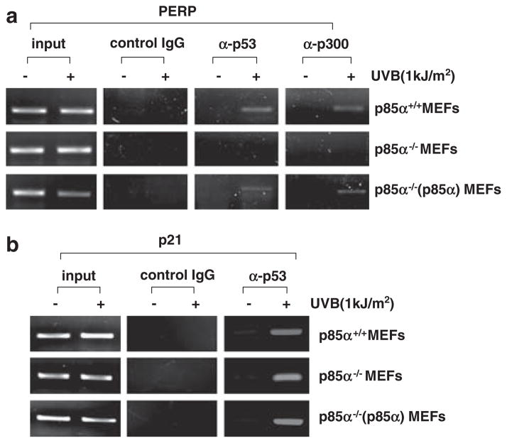
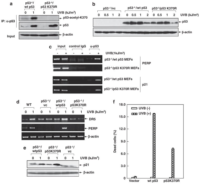
Inducible acetylation of p53 at lysine residues has a great impact on regulating the transactivation of this protein, which is associated with cell growth arrest and/or apoptosis under various stress conditions. However, the factor(s) for regulating p53 acetylation remains largely unknown. In the current study, we have shown that p85α, the regulatory subunit of phosphatidylinositol-3-kinase, has a critical role in mediating p53 acetylation and promoter-specific transactivation in the ultraviolet B (UVB) response. Depletion of p85α in mouse embryonic fibroblasts significantly impairs UVB-induced apoptosis, as well as p53 transactivation and acetylation at Lys370 (Lys373 of human p53); however, the accumulation, nuclear translocation and phosphorylation of p53 are not affected. Interestingly, p85α binds to p300, promotes the p300-p53 interaction and the subsequent recruitment of the p53/p300 complex to the promoter region of the specific p53 target gene in response to UVB irradiation. Moreover, ablation of p53 acetylation at Lys370 by site-directed mutagenesis dramatically suppresses UVB-induced expression of the specific p53-responsive gene as well as cell apoptosis. Therefore, we conclude that p85α is a novel regulator of p53-mediated response under certain stress conditions, and targeting the p85α-dependent p53 pathway may be promising for cancer therapy.


Conflict of interest statement

Conflict of interest

The authors declare no conflict of interest.

Figures

Figure 1
Figure 1
p85α is involved in p53 transactivation induced by UVB irradiation. (a) p85α+/+, p85α−/− and reconstituted p85α−/− (p85α) MEFs were exposed to UVB (1 kJ/m2), and then cell apoptosis was determined by flow cytometric assay at the indicated time points after UVB exposure. (b) p85α+/+ and p85α−/− MEFs with the stable transfection of the p53-luciferase reporter construct were exposed to UVB (1 kJ/m2) for the indicated time periods, and then the p53 luciferase activities were measured. The results were expressed as the relative p53 luciferase activities, which were normalized to the luciferase activity of the cells without any treatment. (c, d) p85α+/+ and p85α−/− MEFs were exposed to the UVB (1 kJ/m2) for the indicated time periods, and then the expression of p53-target genes including DR5, PERP, PUMA, BAX, GADD45α and p21 were detected by semi-quantitative reverse transcription–PCR (RT–PCR) (c) and western blot assays (d). (e) p85α+/+, p85α−/− and p85α−/− (p85α) MEFs with the stable transfection of the p53-luciferase reporter construct were exposed to UVB (1kJ/m2) for the indicated time periods, and then the p53 luciferase activities were measured. (f) p85α+/+, p85α−/− and p85α−/− (p85α) MEFs were exposed to UVB (1 kJ/m2) for the indicated time periods and then the transcriptional induction of PERP and DR5 were detected by semi-quantitative RT–PCR assay. (g) p85α+/+ (control shRNA) and p85α+/+ (p85α shRNA) MEFs were exposed to UVB (1 kJ/m2), and then cell apoptosis was determined by detecting caspase-3 cleavage at the indicated time points after UVB exposure. (h) p85α+/+ (control shRNA) and p85α+/+ (p85α shRNA) MEFs with the stable transfection of the p53-luciferase reporter construct were exposed to UVB (1 kJ/m2) for the indicated time periods, and then the p53 luciferase activities were measured. (i) p85α+/+ (control shRNA) and p85α+/+ (p85α shRNA) MEFs were exposed to UVB (1 kJ/m2) for the indicated time periods, and then the transcriptional induction of PERP and DR5 were detected by semi-quantitative RT–PCR assay.
Figure 2
Figure 2
p85α deficiency does not alter the accumulation, phosphorylation and nuclear translocation of p53 induced by UVB. (a and b) p85α+/+ and p85α−/− MEFs were either exposed to a single dose (1 kJ/m2) of UVB for the indicated time periods (a) or the different doses of UVB for 12 h (b), and then the accumulation and phosphorylation of p53 (Ser18, Ser389) were detected by western blot assay. (c) p85α+/+ and p85α−/− MEFs were exposed to UVB (1 kJ/m2) for 12 h, and then the cytoplasmic and nuclear proteins were extracted and subjected to the western blot assay to determine the nuclear translocation of p53. (d) p85α+/+ (control shRNA) and p85α+/+ (p85α shRNA) MEFs were exposed to UVB (1 kJ/m2) for the indicated time periods, and then the accumulation and phosphorylation of p53 (Ser18, Ser389) were detected as described above.
Figure 3
Figure 3
p85α is required for p53 acetylation at Lys370 in the UVB response. (a) p85α+/+ and p85α−/− MEFs were exposed to UVB (1 kJ/m2) for the indicated time periods, and then whole cell extracts were immunoprecipitated with the anti-p53 antibody. The immunoprecipitated samples were probed with anti-p53-AC-K370 and anti-p53-AC-K379 antibodies to detect the acetylation of p53 at Lys370 and Lys379 in the UVB response. Total p53 levels were detected in the whole cell extract. (b) p85α+/+, p85α−/− and p85α−/− (p85α) MEFs were exposed to UVB (1 kJ/m2) for 8 h, and then the accumulation and acetylation of p53 at Lys370 and Lys379 were detected as described above. (c) p85α+/+ (p85α shRNA) MEFs were left untreated or transiently transfected with p85α expression plasmid. After 36 h, the transfected cells and the p85α+/+ (control shRNA) MEFs were exposed to UVB (1 kJ/m2) for 8 h, and then the accumulation and acetylation of p53 at Lys370 and Lys379 were detected. IP, immunoprecipitation.
Figure 4
Figure 4
p85α interacts with p300, enhances p300–p53 interaction and the autoacetylation of p300 in the cellular response to UVB irradiation. (a) 293T cells were co-transfected with the expression plasmids containing HA-p300 and GFP-p85α or the control vector. After 36 h of transfection, the cells were exposed to UVB as the indicated time periods, and then whole cell lysates were immunoprecipitated with the anti-GFP antibody. The immunoprecipitated samples were then probed with anti-p300 antibody to determine the interaction between p85α and p300 under UVB irradiation. (b) NIH3T3 cell lysates were immunoprecipitated with anti-p300 antibody or control immunoglobulin G (IgG), and then the immunoprecipitated samples were probed with the anti-p85α antibody to detect the binding of endogenous p85α to p300. (ce) The samples described in Figures 3a–c were probed with anti-p300 and anti-p300-AC-Lys1499 antibodies to detect the binding of p300 to p53 and the autoacetylation of p300 under UVB irradiation, respectively. IP, immunoprecipitation.
Figure 5
Figure 5
p85α-dependent p53 acetylation is essential for the promoter-specific binding ability of p53. Soluble chromatin prepared from UVB-treated and -untreated p85α+/+, p85α−/− and p85α−/−(p85α) MEFs was subjected to a ChIP assay using the normal immunoglobulin G, anti-p53 or anti-p300 antibodies, respectively. Immunoprecipitated chromatin DNA was PCR-amplified with the primers that specifically annealed to the region flanking p53-responsive elements within the promoter region of PERP (a) or p21 (b).
Figure 6
Figure 6
p85α-dependent p53 acetylation at Lys370 is critical for promoter-specific p53 transactivation in the UVB response. (a) p53−/− MEFs were transfected with the expression plasmids containing wt p53 or p53K370R. After 36 h of transfection, the cells were exposed to UVB (1 kJ/m2) for 8 h, and the whole cell lysates were immunoprecipitated with the anti-p53 antibody. The immunoprecipitated samples were then probed with anti-p53 or anti-p53-AC-K370 antibodies to detect the accumulation and acetylation (K370) of p53. (b) p53−/− MEFs were transfected with the expression plasmids containing wt p53, the p53K370R or the control vector (vc). After 36 h of transfection, the cells were exposed to different doses of UVB, and then the accumulation of p53 was detected by a western blot assay. (c) p53−/− MEFs were transfected with the wt p53 or p53K370R and then exposed to UVB (1 kJ/m2) for 8 h. The ChIP assay was performed using normal serum or the anti-p53 antibody. Immunoprecipitated chromatin DNA was PCR-amplified with the primers that specifically annealed to the region flanking p53-responsive elements within the PERP or p21 genes. (d) p53−/− MEFs were transfected with the expression plasmids containing wt p53, p53K370R or the control vector. After 36 h of transfection, the cells were exposed to UVB (1 kJ/m2), and the transcription of PERP and DR5 were detected by a reverse transcription PCR assay 12 h after UVB exposure. The same assay performed using WT MEFs was also shown here as a positive control. (e) p53−/− MEFs were transfected and treated as described in (d), and then the induction of p21 expression was determined by western blot assay 12 h after UVB exposure. (f) p53−/− MEFs were transfected and treated as described in (d), and then the apoptosis of the transfected cells was determined by a flow cytometric assay 24 h after UVB exposure. IP, immunoprecipitation.

Similar articles

Cited by

References

    1. An W, Kim J, Roeder RG. Ordered cooperative functions of PRMT1, p300, and CARM1 in transcriptional activation by p53. Cell. 2004;117:735–748. - PubMed
    1. Avantaggiati ML, Ogryzko V, Gardner K, Giordano A, Levine AS, Kelly K. Recruitment of p300/CBP in p53-dependent signal pathways. Cell. 1997;89:1175–1184. - PubMed
    1. Brooks CL, Gu W. Ubiquitination, phosphorylation and acetylation: the molecular basis for p53 regulation. Curr Opin Cell Biol. 2003;15:164–171. - PubMed
    1. Brooks CL, Gu W. p53 activation: a case against Sir. Cancer Cell. 2008;13:377–378. - PMC - PubMed
    1. Cao C, Lu S, Kivlin R, Wallin B, Card E, Bagdasarian A, et al. SIRT1 confers protection against UVB- and H(2)O(2)-induced cell death via modulation of p53 and JNK in cultured skin keratinocytes. J Cell Mol Med. 2008;4:4. - PMC - PubMed

Publication types

Substances