. 2011 Mar;13(1):20-9.
doi: 10.1208/s12248-010-9240-y. Epub 2010 Nov 6.

Microcalorimetric method to assess phagocytosis: macrophage-nanoparticle interactions

Affiliations

Microcalorimetric method to assess phagocytosis: macrophage-nanoparticle interactions

M H D Kamal Al-Hallak et al. AAPS J. 2011 Mar.

Abstract

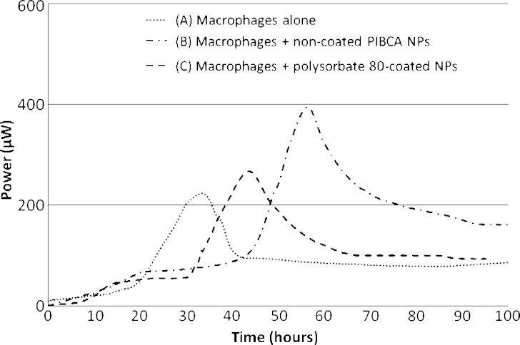
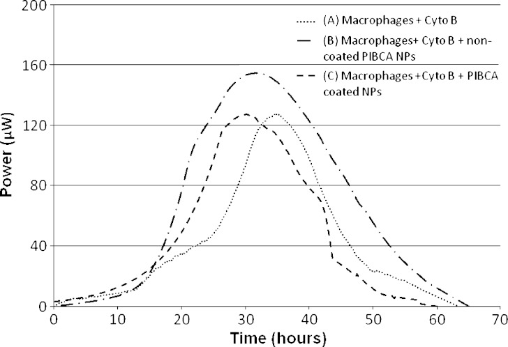
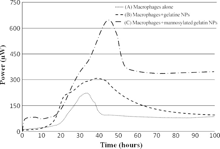
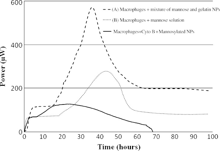
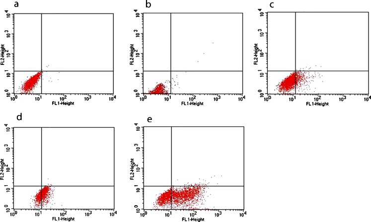
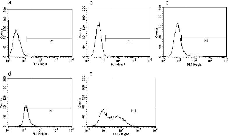
This study evaluated the use of isothermal microcalorimetry (ITMC) to detect macrophage-nanoparticle interactions. Four different nanoparticle (NP) formulations were prepared: uncoated poly(isobutyl cyanoacrylate) (PIBCA), polysorbate-80-coated PIBCA, gelatin, and mannosylated gelatin NPs. Changes in NP formulations were aimed to either enhance or decrease macrophage-NP interactions via phagocytosis. Alveolar macrophages were cultured on glass slabs and inserted in the ITMC instrument. Thermal activities of the macrophages alone and after titration of 100 μL of NP suspensions were compared. The relative interactive coefficients of macrophage-NP interactions were calculated using the heat exchange observed after NP titration. Control experiments were performed using cytochalasin B (Cyto B), a known phagocytosis inhibitor. The results of NP titration showed that the total thermal activity produced by macrophages changed according to the NP formulation. Mannosylated gelatin NPs were associated with the highest heat exchange, 75.4 ± 7.5 J, and thus the highest relative interactive coefficient, 9,269 ± 630 M-1. Polysorbate-80-coated NPs were associated with the lowest heat exchange, 15.2 ± 3.4 J, and the lowest interactive coefficient, 890 ± 120 M-1. Cyto B inhibited macrophage response to NPs, indicating a connection between the thermal activity recorded and NP phagocytosis. These results are in agreement with flow cytometry results. ITMC is a valuable tool to monitor the biological responses to nano-sized dosage forms such as NPs. Since the thermal activity of macrophage-NP interactions differed according to the type of NPs used, ITMC may provide a method to better understand phagocytosis and further the development of colloidal dosage forms.


Figures

Fig. 1
Fig. 1
The thermal activity profiles of A macrophages alone, B macrophages after titration of 100 μL of uncoated poly(isobutyl cyanoacrylat) nanoparticles, and C macrophages after titration of 100 μL of polysorbate-80-coated poly(isobutyl cyanoacrylat) nanoparticles
Fig. 2
Fig. 2
The thermal activity profiles of A macrophages alone, B macrophages after titration of 100 μL of non-coated poly(isobutyl cyanoacrylat) nanoparticles, and C macrophages after titration of 100 μL of polysorbate-80-coated poly(isobutyl cyanoacrylat) nanoparticles after the cytochalasin B was added at the same concentration (2–5 × 10−6 M) to the medium used in all experiment
Fig. 3
Fig. 3
The thermal activity profiles of A macrophages alone, B macrophages after titration of 100 μl of gelatin nanoparticles, and C macrophages after titration of 100 μl of mannosylated gelatin nanoparticles
Fig. 4
Fig. 4
The thermal activity profiles of A macrophages after titration 100 μl of 1:1 mixture of gelatin nanoparticles and mannose, B macrophages after titrating 100 μl of mannosylated solution of 0.1 mg/ml, and C macrophages after titration of 100 μl of mannosylated gelatin nanoparticles with cytochalasin B added to the cell culture medium
Fig. 5
Fig. 5
Flow cytometric dot plots of (a) untreated macrophages, (b) macrophages treated with polysorbate-80-coated PIBCA nanoparticles, (c) macrophages treated with uncoated PIBCA nanoparticles, (d) macrophages treated with gelatin nanoparticles and (e) macrophages treated with mannosylated gelatin nanoparticles
Fig. 6
Fig. 6
Flow cytometric analysis of the macrophage uptake of nanoparticles: (a) represent the macrophages auto-fluorescences MFI = 3.19. the horizontal line was set to calculate the percentage of positive cells, (b) poysorbate-80 coated PIBCA nanoparticles MFI = 5.07, (c) uncoated PIBCA nanoparticles MFI = 16.81, (d) gelatin nanoparticles MFI = 18.39 and (e) mannosylated gelatin nanoparticles MFI = 46.08

Similar articles

Cited by

References

    1. Oh Y-K, Swanson JA. Different fates of phagocytosed particles after delivery into macrophage lysosomes. J Cell Biol. 1996;132(4):585–593. doi: 10.1083/jcb.132.4.585. - DOI - PMC - PubMed
    1. Schafer V, von Briesen H, Rubsamen-Waigmann H, Steffan AM, Royer C, Kreuter J. Phagocytosis and degradation of human serum albumin microspheres and nanoparticles in human macrophages. J Microencapsul. 1994;11(3):261–269. doi: 10.3109/02652049409040455. - DOI - PubMed
    1. Roser M, Fischer D, Kissel T. Surface-modified biodegradable albumin nano- and microspheres. II: effect of surface charges on in vitro phagocytosis and biodistribution in rats. Eur J Pharm Biopharm. 1998;46(3):255–263. doi: 10.1016/S0939-6411(98)00038-1. - DOI - PubMed
    1. Al-Hallak KM, Azarmi S, Anwar-Mohamed A, Roa WH, Lobenberg R. Secondary cytotoxicity mediated by alveolar macrophages: a contribution to the total efficacy of nanoparticles in lung cancer therapy? Eur J Pharm Biopharm. 2010;76(1):112–9. doi: 10.1016/j.ejpb.2010.05.002. - DOI - PubMed
    1. Soma CE, Dubernet C, Barratt G, Benita S, Couvreur P. Investigation of the role of macrophages on the cytotoxicity of doxorubicin and doxorubicin-loaded nanoparticles on M5076 cells in vitro. J Control Release. 2000;68(2):283–289. doi: 10.1016/S0168-3659(00)00269-8. - DOI - PubMed

Publication types

MeSH terms

LinkOut - more resources