Nephrocalcinosis: re-defined in the era of endourology
- PMID: 21057942
- PMCID: PMC3787841
- DOI: 10.1007/s00240-010-0328-8
Nephrocalcinosis: re-defined in the era of endourology
Abstract
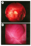
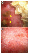
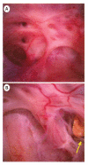
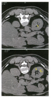
Nephrocalcinosis generally refers to the presence of calcium salts within renal tissue, but this term is also used radiologically in diagnostic imaging in disease states that also produce renal stones, so that it is not always clear whether it is tissue calcifications or urinary calculi that give rise to the characteristic appearance of the kidney on x-ray or computed tomography (CT). Recent advances in endoscopic imaging now allow the visual distinction between stones and papillary nephrocalcinosis, and intrarenal endoscopy can also verify the complete removal of urinary stones, so that subsequent radiographic appearance can be confidently attributed to nephrocalcinosis. This report shows exemplary cases of primary hyperparathyroidism, type I distal renal tubular acidosis, medullary sponge kidney, and common calcium oxalate stone formation. In the first three cases--all being conditions commonly associated with nephrocalcinosis--it is shown that the majority of calcifications seen by radiograph may actually be stones. In common calcium oxalate stones formers, it is shown that Randall's plaque can appear as a small calculus on CT scan, even when calyces are known to be completely clear of stones. In the current era with the use of non-contrast CT for the diagnosis of nephrolithiasis, the finding of calcifications in close association with the renal papillae is common. Distinguishing nephrolithiasis from nephrocalcinosis requires direct visual inspection of the papillae and so the diagnosis of nephrocalcinosis is essentially an endoscopic, not radiologic, diagnosis.
Figures


References
-
- Albright F, Baird PC, Cope O, Bloomberg E. Studies on the physiology of the parathyroid glands. IV. Renal complications of hyperparathyroidism. Am J Med Sci. 1934;187(1):49–64.
-
- Engel WJ. Urinary calculi associated with nephrocalcinosis. J Urol. 1952;68(1):105–116. - PubMed
-
- Spence HM, Singleton R. What is sponge kidney disease and where does it fit in the spectrum of cystic disorders? J Urol. 1972;107(2):176–183. - PubMed
-
- Mortensen JD, Emmett JL. Nephrocalcinosis; a collective and clinicopathologic study. J Urol. 1954;71(4):398–406. - PubMed
-
- Evan AP, Lingeman J, Coe FL, Worcester E. Randall's plaque: pathogenesis and role in calcium oxalate nephrolithiasis. Kidney Int. 2006;69:1313–1318. - PubMed
Publication types
MeSH terms
Substances
Grants and funding
LinkOut - more resources
Full Text Sources
Other Literature Sources

