Review
. 2011 Jan;121(1):79-90.
doi: 10.1007/s00401-010-0761-3. Epub 2010 Nov 7.

Molecular biology and pathology of prion strains in sporadic human prion diseases

Affiliations
Review

Molecular biology and pathology of prion strains in sporadic human prion diseases

Pierluigi Gambetti et al. Acta Neuropathol. 2011 Jan.

Abstract

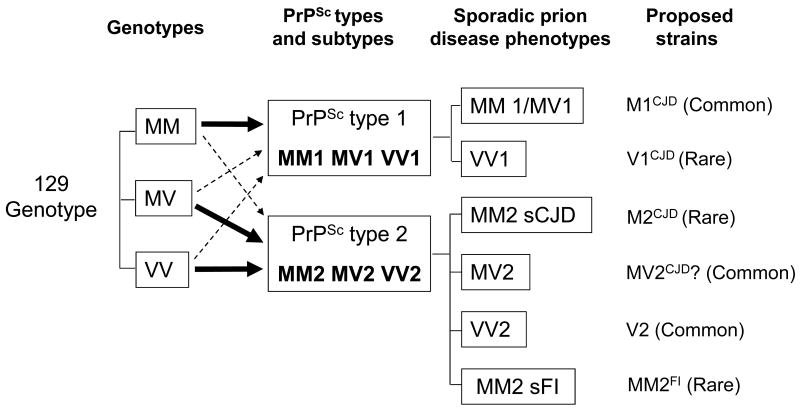
Prion diseases are believed to propagate by the mechanism involving self-perpetuating conformational conversion of the normal form of the prion protein, PrP(C), to the misfolded, pathogenic state, PrP(Sc). One of the most intriguing aspects of these disorders is the phenomenon of prion strains. It is believed that strain properties are fully encoded in distinct conformations of PrP(Sc). Strains are of practical relevance to human prion diseases as their diversity may explain the unusual heterogeneity of these disorders. The first insight into the molecular mechanisms underlying heterogeneity of human prion diseases was provided by the observation that two distinct disease phenotypes and their associated PrP(Sc) conformers co-distribute with distinct PrP genotypes as determined by the methionine/valine polymorphism at codon 129 of the PrP gene. Subsequent studies identified six possible combinations of the three genotypes (determined by the polymorphic codon 129) and two common PrP(Sc) conformers (named types 1 and 2) as the major determinants of the phenotype in sporadic human prion diseases. This scenario implies that each 129 genotype-PrP(Sc) type combination would be associated with a distinct disease phenotype and prion strain. However, notable exceptions have been found. For example, two genotype-PrP(Sc) type combinations are linked to the same phenotype, and conversely, the same combination was found to be associated with two distinct phenotypes. Furthermore, in some cases, PrP(Sc) conformers naturally associated with distinct phenotypes appear, upon transmission, to lose their phenotype-determining strain characteristics. Currently it seems safe to assume that typical sporadic prion diseases are associated with at least six distinct prion strains. However, the intrinsic characteristics that distinguish at least four of these strains remain to be identified.


Figures

Figure 1
Figure 1. Representation of the original identification of two PrPSc conformers as distinct strains subsequently named types 1 and 2
The two PrPSc conformers, referred to as types 2 and 1, were respectively associated with fatal familial insomnia (FFI) and Creutzfeldt-Jakob disease (fCJDD178N) (A), two familial prion diseases that share the same D178N mutation in the PrP gene but have distinct phenotypes as demonstrated by the opposite topography of the lesions in thalamus and cerebral cortex (B). PrPSc types 2 and 1 also show distinct electrophoretic profiles (C), which can be appreciated by the distinct mobilities to kDa 19 and kDa 21 of the lowest electrophoretic bands corresponding to the unglycosylated fragments of PrP resistant to protease digestion. The different electrophoretic mobilities of the two PrPSc conformers support the notion that they have different conformations represented as different geometric forms (C). PrPSc types 2 and 1 associated with FFI and fCJDE200K another form of familial CJD was replicated upon inoculation to receptive mice (D), even in the absence of the original PrP gene mutation. Familial CJDD178N could not be transmitted to the same transgenic mice probably because 129M/129V mismatch. formula image
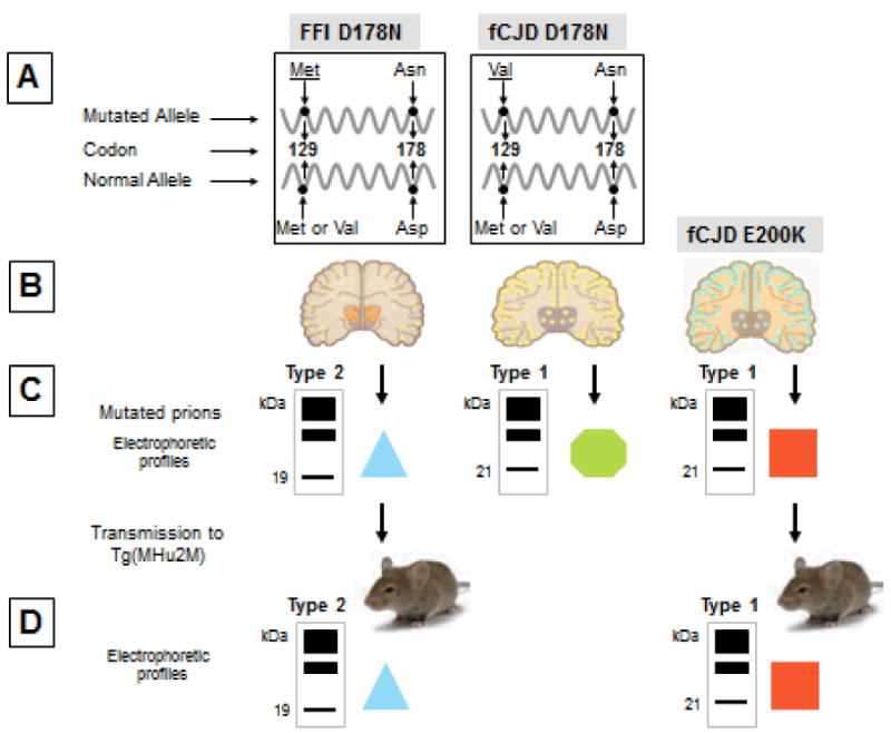
: The light blue triangle represents the conformation of the PrPSc conformer from FFID178N; formula image: The green hexagon represents the conformation of PrPSc from fCJDD178N; formula image: The red square represents the conformation of PrPSc from fCJDE200K which although being type one as fCJDD178N- associated PrPSc is presumably different based on the fCJDE200K distinct phenotype.
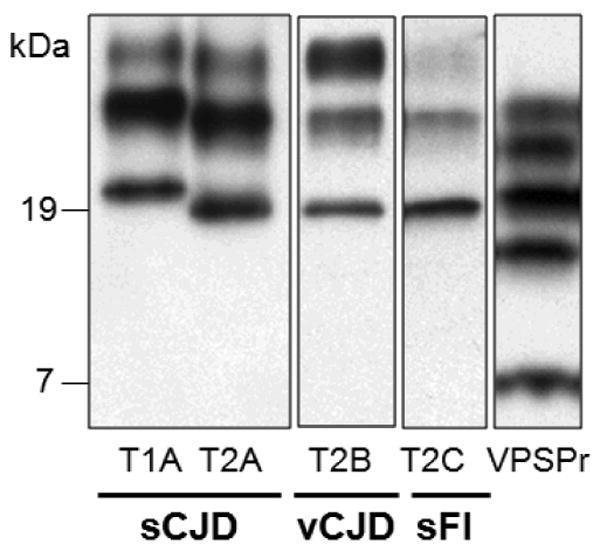
Figure 2
Figure 2. Western blots of PrPSc types 1 and 2, and of PrPDis associated with VPSPr-129MM
The different electrophoretic mobilities of the PrPSc types 1 (T1A) and 2 (T2A-C) are evident. The capital letters identify the glycoform ratios that may occur in PrPSc types 1 and 2. The overrepresentation of the middle band containing the monoglycosylated isoform is indicated with A; B indicates the overrepresentation of the upper band (diglycosylated isoform) and C that of the lowest band (unglycosylated isoform). The ladder-like profile formed by the PrPDis indicated as VPSPr is from VPSPr-129MM but this profile is shared by the three VPSPr 129 genotypes. All preparations have been treated with proteinase K. The sporadic Creutzfeldt-Jakob disease (sCJD) and variant (v) CJD preparations have been probed with 3F4, while the sporadic fatal insomnia (sFI) and the variably protease-sensitive prionopathy (VPSPr) preparations have been probed with IE4, both monoclonal antibodies to the prion protein.
Figure 3
Figure 3. Diagram of the cleavage pattern of PrPSc types 1 and 2 associated with the three 129 genotypes
The large arrowheads indicate the major sites of cleavage by proteinase K, which are further identified with the residue numbers of the prion protein (PrP) (lowest bar). The smaller arrowheads indicate the minor cleavage sites. Minor cleavage sites extend more towards the C-terminus in PrPSc type 1, while they extend more toward the N-terminus in at least 2 of the PrPSc type 2 preparations (MV2 and VV2). This phenomenon is more prominent when PrPSc types 1 and 2 are associated with the 129MV genotype indicating that in this association, PrPSc 1 and 2 N-termini are more ragged.
Figure 4
Figure 4. Conformation stability immunoassay (CSI) of the unglycosylated isoform of PrPSc from sCJDMM1, sCJDMM2 and sCJDMM1-2
To measure the stability of PrPSc, increasing concentrations of GdnHCl are used to unfold PrPSc, which in turn becomes progressively more sensitive to digestion with PK. The concentration of guanidine applied to achieve the digestion of 50% of the PrPSc with proteinase K (PK) is indicated by [GdnHCl]1/2 (shown by the vertical color coded broken lines). The CSI curve shows that the stability of the PrPSc type 1 when it is present alone as in sCJDMM1 (red hollow diamond) is significantly different from the stability that it shows when it co-occurs with PrPSc type 2 as in sCJDMM1-2 (green solid triangle) ([GdnHCl]1/2 values 2.96M vs. 1.96M, respectively; p<10-5). Furthermore, the CSI curve of the PrPSc type 1 from sCJDMM1-2 becomes indistinguishable from that of PrPSc type 2 from sCJDMM2 (black square with hollow circle) and sCJDMM1-2 (blue solid circle), which are similar indicating no change in CSI characteristics between PrPSc 2 when alone or co-occurring with type 1. CSI profiles are smooth curves best matching data (mean ± SD) obtained at different concentrations of guanidine.
Figure 5
Figure 5. Proposed relationships between the 129 genotypes, PrPSc types and subtypes, phenotypes and strains in sporadic Creutzfeldt-Jakob (sCJD) and sporadic fatal insomnia (sFI)
The three 129 genotypes (and possibly other polymorphisms) are believed to influence the formation of PrPSc type 1 or 2 as the 129MM genotype is preferentially associated with type 1 while 129MV and 129VV are associated with type 2. The six possible combinations of the three 129 genotypes with PrPSc types 1 and 2 are associated with six distinct disease phenotypes, five belonging to sCJD and one to sFI. The pairing of 129 genotype-PrPSc type combinations with the phenotypes is not stringent. Notable exceptions are the association of the distinct MM1 and MV1 combinations with one phenotype (MM1/MV1) and the association of the single MM2 combination with two phenotypes (MM2 sCJD and MM2 sFI). It is proposed that the six phenotypes are paired with six distinct prion strains, three of which are relatively prevalent, while three are rare and might result from variations in the strain selection process. The distinct strain proposed to be associated with sCJDMV2 is questionable because of the inability to maintain its distinctive characteristics after transmission [5]. Coexistence of types 1 and 2 associated with the three 129 genotypes and the strains possibly associated with the novel disease variably protease-sensitive prionopathy (VPSPr), are not shown for simplicity, and because the presence of true strains in VPSPr is currently unclear.

References

    1. Angers RC, Kang HE, Napier D, et al. Prion strain mutation determined by prion protein conformational compatibility and primary structure. Science. 2010;328:1154–1158. - PMC - PubMed
    1. Apetri AC, Vanik DL, Surewicz WK. Polymorphism at residue 129 modulates the conformational conversion of the D178N variant of human prion protein 90-231. Biochemistry. 2005;44:15880–15888. - PubMed
    1. Apostol MI, Sawaya MR, Cascio D, Eisenberg D. Crystallographic studies of PrP segments suggest how structural changes encoded by polymorphism at residue 129 modulate susceptibility to human prion disease. J Biol Chem. 2010 Aug 4; Epub ahead of print. - PMC - PubMed
    1. Bessen RA, Marsh RF. Identification of two biologically distinct strains of transmissible mink encephalopathy in hamsters. J Gen Virol. 1992;73:329–334. - PubMed
    1. Bishop MT, Will RG, Manson JC. Defining sporadic Creutzfeldt-Jakob disease strains and their transmission properties. Proc Natl Acad Sci U S A. 2010;107:12005–12010. - PMC - PubMed

Publication types