

Absorption and translocation to the aerial part of magnetic carbon-coated nanoparticles through the root of different crop plants
- PMID: 21059206
- PMCID: PMC2994779
- DOI: 10.1186/1477-3155-8-26
Absorption and translocation to the aerial part of magnetic carbon-coated nanoparticles through the root of different crop plants
Abstract
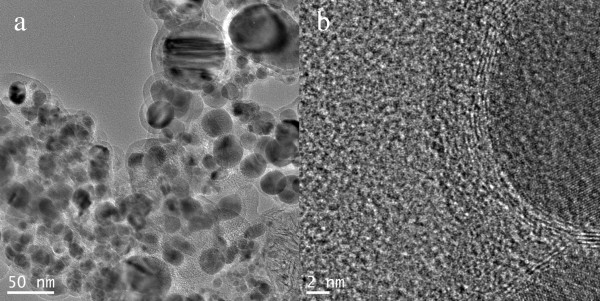
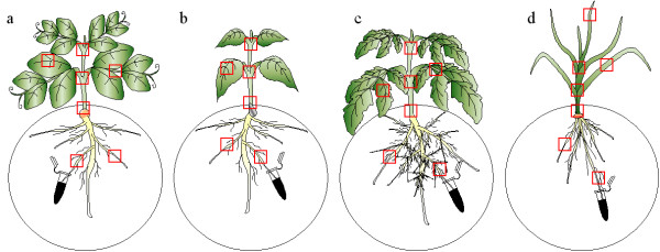
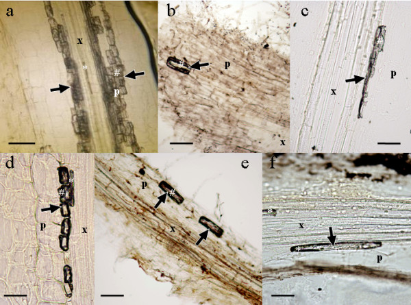
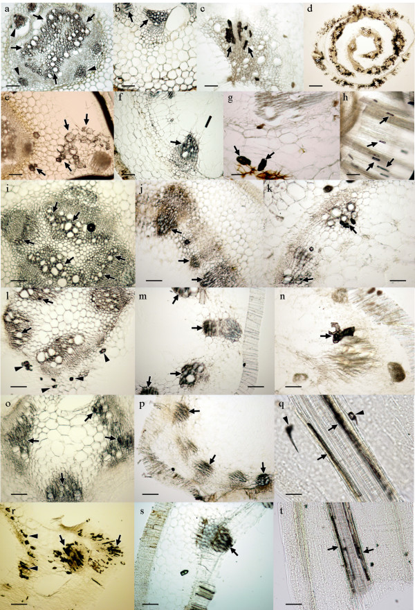
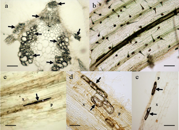
The development of nanodevices for agriculture and plant research will allow several new applications, ranging from treatments with agrochemicals to delivery of nucleic acids for genetic transformation. But a long way for research is still in front of us until such nanodevices could be widely used. Their behaviour inside the plants is not yet well known and the putative toxic effects for both, the plants directly exposed and/or the animals and humans, if the nanodevices reach the food chain, remain uncertain. In this work we show that magnetic carbon-coated nanoparticles forming a biocompatible magnetic fluid (bioferrofluid) can easily penetrate through the root in four different crop plants (pea, sunflower, tomato and wheat). They reach the vascular cylinder, move using the transpiration stream in the xylem vessels and spread through the aerial part of the plants in less than 24 hours. Accumulation of nanoparticles was detected in wheat leaf trichomes, suggesting a way for excretion/detoxification. This kind of studies is of great interest in order to unveil the movement and accumulation of nanoparticles in plant tissues for assessing further applications in the field or laboratory.
Figures
Similar articles
-
Nanoparticles as smart treatment-delivery systems in plants: assessment of different techniques of microscopy for their visualization in plant tissues.Ann Bot. 2008 Jan;101(1):187-95. doi: 10.1093/aob/mcm283. Epub 2007 Nov 11. Ann Bot. 2008. PMID: 17998213 Free PMC article.
-
Carbon-iron magnetic nanoparticles for agronomic use in plants: promising but still a long way to go.Plant Signal Behav. 2010 Oct;5(10):1295-7. doi: 10.4161/psb.5.10.13080. Epub 2010 Oct 1. Plant Signal Behav. 2010. PMID: 20930509 Free PMC article.
-
Barriers, pathways and processes for uptake, translocation and accumulation of nanomaterials in plants--Critical review.Nanotoxicology. 2016;10(3):257-78. doi: 10.3109/17435390.2015.1048326. Epub 2015 Jun 11. Nanotoxicology. 2016. PMID: 26067571 Review.
-
Synthesis, application, and tracking of magnetic carbon-coated nanoparticles in plants.Methods Mol Biol. 2012;906:263-72. doi: 10.1007/978-1-61779-953-2_20. Methods Mol Biol. 2012. PMID: 22791439
-
Arsenic behavior in soil-plant system and its detoxification mechanisms in plants: A review.Environ Pollut. 2021 Oct 1;286:117389. doi: 10.1016/j.envpol.2021.117389. Epub 2021 May 21. Environ Pollut. 2021. PMID: 34058445 Review.
Cited by
-
Effects of Gold Nanoparticles on Mentha spicata L., Soil Microbiota, and Human Health Risks: Impact of Exposure Routes.Nanomaterials (Basel). 2024 May 29;14(11):955. doi: 10.3390/nano14110955. Nanomaterials (Basel). 2024. PMID: 38869580 Free PMC article.
-
Impact of Zinc oxide nanoparticles on eggplant (S. melongena): studies on growth and the accumulation of nanoparticles.IET Nanobiotechnol. 2018 Sep;12(6):706-713. doi: 10.1049/iet-nbt.2017.0237. IET Nanobiotechnol. 2018. PMID: 30104442 Free PMC article.
-
Reducing Nitrogen Dosage in Triticum durum Plants with Urea-Doped Nanofertilizers.Nanomaterials (Basel). 2020 May 29;10(6):1043. doi: 10.3390/nano10061043. Nanomaterials (Basel). 2020. PMID: 32486000 Free PMC article.
-
Penetration and Toxicity of Nanomaterials in Higher Plants.Nanomaterials (Basel). 2015 May 26;5(2):851-873. doi: 10.3390/nano5020851. Nanomaterials (Basel). 2015. PMID: 28347040 Free PMC article. Review.
-
Vitamin B12 (Cobalamin) and Micronutrient Fortification in Food Crops Using Nanoparticle Technology.Front Plant Sci. 2021 Aug 23;12:668819. doi: 10.3389/fpls.2021.668819. eCollection 2021. Front Plant Sci. 2021. PMID: 34497618 Free PMC article. Review.
References
-
- Robinson DKR, Morrison M. Nanotechnology Developments for the Agrifood sector- Report of the ObservatoryNANO. Institute of Nanotechnology, UK; 2009. http://observatorynano.eu/
LinkOut - more resources
Full Text Sources
