. 2011 Jan 14;286(2):1649-58.
doi: 10.1074/jbc.M110.106922. Epub 2010 Nov 8.

Aberrant estrogen regulation of PEMT results in choline deficiency-associated liver dysfunction

Affiliations

Aberrant estrogen regulation of PEMT results in choline deficiency-associated liver dysfunction

Mary E Resseguie et al. J Biol Chem. .

Abstract

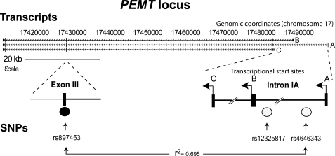
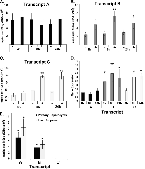
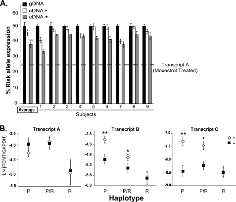
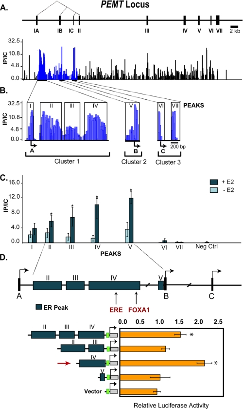
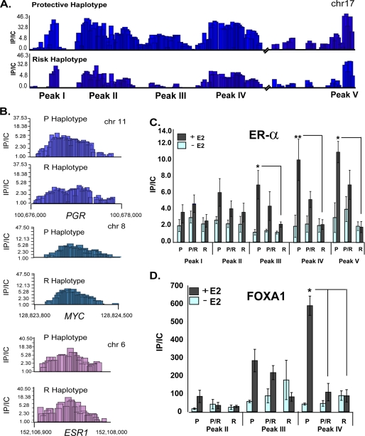
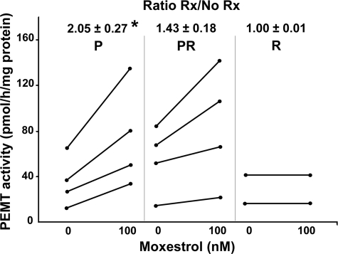
When dietary choline is restricted, most men and postmenopausal women develop multiorgan dysfunction marked by hepatic steatosis (choline deficiency syndrome (CDS)). However, a significant subset of premenopausal women is protected from CDS. Because hepatic PEMT (phosphatidylethanolamine N-methyltransferase) catalyzes de novo biosynthesis of choline and this gene is under estrogenic control, we hypothesized that there are SNPs in PEMT that disrupt the hormonal regulation of PEMT and thereby put women at risk for CDS. In this study, we performed transcript-specific gene expression analysis, which revealed that estrogen regulates PEMT in an isoform-specific fashion. Locus-wide SNP analysis identified a risk-associated haplotype that was selectively associated with loss of hormonal activation. Chromatin immunoprecipitation, analyzed by locus-wide microarray studies, comprehensively identified regions of estrogen receptor binding in PEMT. The polymorphism (rs12325817) most highly linked with the development of CDS (p < 0.00006) was located within 1 kb of the critical estrogen response element. The risk allele failed to bind either the estrogen receptor or the pioneer factor FOXA1. These data demonstrate that allele-specific ablation of estrogen receptor-DNA interaction in the PEMT locus prevents hormone-inducible PEMT expression, conferring risk of CDS in women.


Figures

FIGURE 1.
FIGURE 1.
PEMT SNPs are associated with CDS risk in women. Shown is a schematic representation of the PEMT locus, including alternative transcriptional start sites (A, B, and C), and location of SNPs (rs12325817 and rs4646343) highly linked to the CDS phenotype in humans fed a low choline diet. Using HapMap data, other SNPs in this region were found to be in high linkage disequilibrium with rs4646343, including an SNP located in common exon 3 (rs897453) (Centre d'Etude du Polymorphisme Humain (CEPH); r2 = 0.695).
FIGURE 2.
FIGURE 2.
Estrogen regulation of PEMT is transcript-specific. Using RNA isolated from primary hepatocytes cultured in the absence (−) or presence (+) of 100 nmol/liter moxestrol for the lengths of time noted, PEMT transcripts were quantified using transcript-specific primers by quantitative PCR. A, transcript A, initially the most abundant, was not responsive to hormone treatment. B, transcript B was significantly induced in response to hormone treatment at both 8 and 24 h after treatment. C, transcript C was the least abundant but was significantly induced in response to hormone treatment at 8 and 24 h. D, PEMT expression levels, presented as a ratio of treated/untreated for all transcripts (normalized to GAPDH), demonstrated significant induction of B (all time points tested) and transcript C (at the 8 and 24 h time points). E, RNA isolated from male human liver biopsies and primary human hepatocytes was subjected to transcript-specific quantitative PCR. For human biopsy samples, results are presented as an average for all subjects (n = 6), and for human hepatocytes, results are shown as an average of all untreated samples, across all time points. Transcripts A and B were significantly more abundant than transcript C. Transcript A was significantly more abundant than transcript B (n = 5–6/time point; *, p < 0.05; **, p < 0.01). Error bars, S.E.
FIGURE 3.
FIGURE 3.
PEMT risk allele is not estrogen-responsive. A, RNA isolated from primary hepatocytes heterozygous for the risk allele was subjected to allele-specific quantitative PCR. Results were normalized to genomic DNA (black bars). The estimated relative expression of the risk allele cDNA versus the protective allele cDNA was calculated by linear regression analysis. In the absence of 100 nmol/liter moxestrol treatment (−, open bars), the risk allele was underexpressed by ∼4% relative to the protective allele. Upon moxesterol treatment (24 h) (+, gray bars), the risk allele was underexpressed by ∼8%. Based on the transcript-specific expression data, the estimated relative abundance of unregulated transcript A in treated cells is depicted by the dashed line. Results are presented for individual subjects and as an average for all subjects (n = 9). *, p < 0.01; **, p < 0.001. Error bars, S.E. B, RNA was isolated from moxestrol-treated (open diamonds) or untreated hepatocytes (black squares) that were P, R, or P/R. PEMT levels were determined by qPCR. Transcript A was not induced by estrogen in any of the groups. Transcripts B and C were induced by hormone in the P and P/R groups, but in hepatocytes homozygous for the risk allele (R), these transcripts were not induced by hormone treatment. Results are presented as the natural log (LN) of the ratio of PEMT relative to GAPDH gene expression levels in moxestrol-treated versus untreated hepatocytes. (n = 5–6/genotype). *, p < 0.001; **, p < 0.0001. Error bars, S.E.
FIGURE 4.
FIGURE 4.
Identification of ER binding sites within PEMT. The PEMT gene encodes three isoforms that are distinguished by unique leading exons, IA, IB, and IC. The dotted lines indicate splicing of exons IA–IC to common exons II–VII. Chromatin isolated from human hepatocytes incubated for 45 min with 100 nmol/liter estradiol (E2) was immunoprecipitated with anti-ERα antibody and hybridized, together with unfractionated chromatin, to a microarray that densely tiled virtually the entire PEMT locus. A, schematic depiction of the PEMT locus. Locus-wide chromatin immunoprecipitation data are expressed as the ratio of immunoprecipitated chromatin to input control (IP/IC). Only positive enrichment ratios of ChIP signal to input control signal are shown. B, seven ER binding regions were mapped within three clusters in close proximity to transcription start sites A, B, and C. The boxed regions indicate identified peaks. C, quantitative PCR was performed on chromatin from cells before and after 45-min exposure to 100 nmol/liter E2 treatment with primers specific to the seven microarray-defined regions (as well as a negative control (neg ctrl)). ER binding to peaks within intron IA and 3′ to the B TSS was significantly increased by E2 (n = 3). *, p < 0.05. D, ER binding regions (peaks II–IV and peak V) from the protective haplotype were cloned upstream of the SV40 promoter and transfected into primary human hepatocytes grown in hormone-depleted media. Cells were either untreated or treated with 100 nmol/liter moxestrol (MOX). Peak IV (indicated by the red arrow) harbors the consensus ERE. The green box represents the SV40 promoter, which drives expression of the luciferase gene. The data represent the average relative luciferase activity normalized for Renilla luciferase and are expressed as -fold induction relative to the activity in the absence of moxestrol. Absolute values for both treated (+) and untreated (−) cells are presented separately in the table. *, p < 0.05 compared with untreated.
FIGURE 5.
FIGURE 5.
Choline deficiency syndrome-associated risk allele fails to bind ER. A, chromatin isolated from 100 nmol/liter estradiol-treated hepatocytes homozygous for the protective or risk alleles was immunoprecipitated with anti-ERα antibodies and hybridized to densely tiled oligonucleotide microarrays. Estrogen receptor complex (ER) binding was decreased for the risk haplotype at peaks II–IV. Chromatin from estradiol-treated (+) or untreated (−) hepatocytes homozygous (P and R) or heterozygous (P/R) for the protective or risk alleles was immunoprecipitated with anti-ERα (B) and anti-FOXA1 (C) antibodies followed by amplification with primers specific for ER-associated regions. Whereas ER binding to peaks II–IV was significantly induced by estrogen for the protective allele, estrogen failed to significantly augment ER binding to the risk allele (n = 5–6/genotype). *, p = 0.0001. Data are expressed as the ratio of immunoprecipitated chromatin to input control (IP/IC). Error bars, S.E. D, ER binding to representative sites on control genes (PGR, ESR1, and MYC) was unaffected by haplotype.
FIGURE 6.
FIGURE 6.
Less PEMT activity is induced by estrogen in human hepatocytes with the risk allele. Primary human hepatocytes that were P, R, or P/R were treated with 0 or 100 nmol/liter moxestrol for 24 h. PEMT activity was measured and reported as absolute values as well as the -fold increase in activity as compared with the untreated cells (mean ± S.D.). The P group (n = 4) had significantly more activity than the P/R (n = 4) and R (n = 2) groups separately or combined (*, p < 0.05). Rx, treatment with moxestrol.

References

    1. Fischer L. M., daCosta K. A., Kwock L., Stewart P. W., Lu T. S., Stabler S. P., Allen R. H., Zeisel S. H. (2007) Am. J. Clin. Nutr. 85, 1275–1285 - PMC - PubMed
    1. Li Z., Agellon L. B., Vance D. E. (2005) J. Biol. Chem. 280, 37798–37802 - PubMed
    1. Buchman A. L., Ament M. E., Sohel M., Dubin M., Jenden D. J., Roch M., Pownall H., Farley W., Awal M., Ahn C. (2001) JPEN J. Parenter. Enteral Nutr. 25, 260–268 - PubMed
    1. Zeisel S. H., Da Costa K. A., Franklin P. D., Alexander E. A., Lamont J. T., Sheard N. F., Beiser A. (1991) FASEB J. 5, 2093–2098 - PubMed
    1. Bremer J., Greenberg D. M. (1961) Biochim. Biophys. Acta 46, 205–216

Publication types

MeSH terms