. 2011 Feb;25(2):703-13.
doi: 10.1096/fj.10-171488. Epub 2010 Nov 8.

Endothelial CYP epoxygenase overexpression and soluble epoxide hydrolase disruption attenuate acute vascular inflammatory responses in mice

Affiliations

Endothelial CYP epoxygenase overexpression and soluble epoxide hydrolase disruption attenuate acute vascular inflammatory responses in mice

Yangmei Deng et al. FASEB J. 2011 Feb.

Abstract

Cytochrome P-450 (CYP)-derived epoxyeicosatrienoic acids (EETs) possess potent anti-inflammatory effects in vitro. However, the effect of increased CYP-mediated EET biosynthesis and decreased soluble epoxide hydrolase (sEH, Ephx2)-mediated EET hydrolysis on vascular inflammation in vivo has not been rigorously investigated. Consequently, we characterized acute vascular inflammatory responses to endotoxin in transgenic mice with endothelial expression of the human CYP2J2 and CYP2C8 epoxygenases and mice with targeted disruption of Ephx2. Compared to wild-type controls, CYP2J2 transgenic, CYP2C8 transgenic, and Ephx2(-/-) mice each exhibited a significant attenuation of endotoxin-induced activation of nuclear factor (NF)-κB signaling, cellular adhesion molecule, chemokine and cytokine expression, and neutrophil infiltration in lung in vivo. Furthermore, attenuation of endotoxin-induced NF-κB activation and cellular adhesion molecule and chemokine expression was observed in primary pulmonary endothelial cells isolated from CYP2J2 and CYP2C8 transgenic mice. This attenuation was inhibited by a putative EET receptor antagonist and CYP epoxygenase inhibitor, directly implicating CYP epoxygenase-derived EETs with the observed anti-inflammatory phenotype. Collectively, these data demonstrate that potentiation of the CYP epoxygenase pathway by either increased endothelial EET biosynthesis or globally decreased EET hydrolysis attenuates NF-κB-dependent vascular inflammatory responses in vivo and may serve as a viable anti-inflammatory therapeutic strategy.


Figures

Figure 1.
Figure 1.
Baseline characterization of Tie2-CYP2J2-Tr and Tie2-CYP2C8-Tr mice. A) Human CYP2J2 and CYP2C8 mRNA levels (assessed by quantitative RT-PCR) and protein levels (assessed by immunoblotting) were abundant in primary lung endothelial cells (ECs) isolated from Tr mice, but not wild-type (WT) littermates. B) After stimulation with A23187, concentrations of 11,12- and 14,15-EET and DHET (the stable EET metabolite), and the regioisomer sum total, released into primary lung EC medium are significantly higher in Tr mice compared to WT littermates (n=3 isolations/genotype group). *P < 0.05 vs. WT. C, F) Human CYP2J2 (C) and CYP2C8 (F) mRNA levels in lung homogenates are significantly higher in Tr mice from multiple founder lines compared to WT littermates (n=11–12 mice/genotype group). *P < 0.001 vs. WT. D, G) Representative immunoblot of microsomal fractions isolated from whole-lung homogenates demonstrate higher CYP2J2 (D) and CYP2C8 (G) protein expression in Tr mice from multiple founder lines (18) compared to WT littermates. Densitometry analysis demonstrated 1.4 ± 0.1- and 1.6 ± 0.2-fold higher immunoreactivity to anti-CYP2J2pep3 and anti-CYP2C8, respectively, in Tr compared to WT mice. Recombinant CYP2J2 and CYP2C8 protein were included as positive controls and exhibited a slightly higher molecular mass compared to the endogenous protein, consistent with prior reports (30, 31). E, H) CYP2J2 (E) and CYP2C8 (H) immunostaining was completed using the anti-CYP2J2pep1 and anti-CYP2C8 antibodies, respectively. Images are from representative lung sections of Tr and WT littermate control mice. Arrows identify endothelial cell staining in Tr mice, but not WT littermates. No immunostaining was observed with normal rabbit serum (not shown).
Figure 2.
Figure 2.
Baseline characterization of Ephx2−/− mice. A, B) Expression of murine sEH protein (assessed by immunoblotting) in primary lung endothelial cells (ECs) (A) and whole lung homogenates (B) was absent in Ephx2−/− mice, but not wild-type (WT) littermates. C) Plasma concentrations of 11,12- and 14,15-EET, and the regioisomer sum total, are significantly higher in Ephx2−/− mice compared to WT littermates (n=7–8/genotype group). *P < 0.01 vs. WT.
Figure 3.
Figure 3.
Endothelial activation by LPS in vitro. A, B) In primary lung endothelial cells (ECs), E-selectin (A) and MCP-1 mRNA (B) levels were increased by LPS (1, 10, 100 ng/ml) in a dose-dependent manner and significantly attenuated in Tie2-CYP2J2-Tr and Tie2-CYP2C8-Tr mice compared to wild-type (WT) littermate controls. C–F) E-selectin (C, E) and MCP-1 (D, F) mRNA and protein levels were also quantified in primary ECs isolated from Tie2-CYP2J2-Tr mice and WT littermate controls (n=3/group) after stimulation with LPS (100 ng/ml) in the absence (vehicle) and presence of 14,15-EEZE (10 μM) or MS-PPOH (10 μM). mRNA data (A–D) were normalized to GAPDH using the 2−ΔCt method. Protein data (E, F) were expressed relative to the saline-treated WT control group. G) Representative immunoblot evaluating phosphorylated IκBα immunoreactivity in EC lysates from the same experiment. H) Densitometry analysis of phosphorylated IκBα immunoreactivity was normalized to GAPDH and expressed relative to the saline-treated WT control group (n=3/group). All data are presented as means ± se. *P < 0.05 vs. WT within same treatment group; #P < 0.05 vs. vehicle within same genotype group.
Figure 4.
Figure 4.
Induction of cytokine, chemokine, and cellular adhesion molecule mRNA expression in vivo. IL-6 and IL-1β (A), MCP-1 and ENA-78 (B), and E-selectin (C) mRNA levels were quantified in lung homogenates 3 h after LPS or saline administration by quantitative RT-PCR. Expression was significantly attenuated in LPS-treated Tie2-CYP2J2-Tr, Tie2-CYP2C8-Tr, and Ephx2−/− mice (n=15–18/group) compared to wild-type (WT) littermates (n=35). Data are presented as mean ± se-fold change in expression, relative to the WT-saline control group (n=21), using the 2−ΔΔCt method. No differences were observed across genotype groups in saline-treated mice (n=4–6/group, data not shown). *P < 0.05 vs. LPS-treated WT group.
Figure 5.
Figure 5.
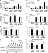
Induction of proinflammatory mediator protein expression and nuclear factor-κB activation in vivo. A, B) E-selectin (A) and MCP-1 (B) protein levels were quantified in lung homogenates 3 h after LPS or saline administration by ELISA. Expression was significantly attenuated in LPS-treated Tie2-CYP2J2-Tr, Tie2-CYP2C8-Tr, and Ephx2−/− mice compared to wild-type (WT) littermates (n=8–10/group). C) Representative immunoblot evaluating IκBα, phosphorylated IκBα, and GAPDH immunoreactivity in lung homogenates 3 h after saline or LPS administration. D, E) Densitometry analysis of phosphorylated IκBα immunoreactivity was normalized to GAPDH (D) and IκBα (E). Each ratio was expressed relative to saline-treated WT controls (n=10), and was lower in Tie2-CYP2J2-Tr, Tie2-CYP2C8-Tr, and Ephx2−/− mice compared to WT littermates (n=6–9/group). Data are presented as means ± se. *P < 0.05 vs. LPS-treated WT group.
Figure 6.
Figure 6.
Histopathological and functional assessment of neutrophil infiltration in vivo. A, B) Semiquantitative score evaluating neutrophil infiltration on H&E-stained (A) and myeloperoxidase (MPO)-immunostained (B) lung sections. All saline-treated wild-type (WT) mice had a score of 0 (n=5). A lower score was observed in LPS-treated Tie2-CYP2J2-Tr, Tie2-CYP2C8-Tr, and Ephx2−/− mice compared to WT littermates (n=5–7/group). C) MPO activity in lung homogenates, expressed relative to saline-treated WT controls (n=12), was significantly lower in LPS-treated Tie2-CYP2J2-Tr, Tie2-CYP2C8-Tr, and Ephx2−/− mice compared to WT littermates (n=10–14/group). Data are presented as means ± se. *P < 0.05 vs. LPS-treated WT group. D–H) Representative images of MPO-immunostained lung sections (×20) in saline-treated WT (D) and LPS-treated WT (E), Tie2-CYP2J2-Tr (F), Tie2-CYP2C8-Tr (G), and Ephx2−/− (H) mice. Arrows indicate MPO staining. Although the saline-treated WT image demonstrates margination of 3 neutrophils within the vessel, this image was selected to confirm the presence of positive immunostaining on the slide. The vast majority of vessels in saline-treated WT mice demonstrated no evidence of neutrophil margination. Representative images of H&E-stained sections are provided in Supplemental Fig. S1.

References

    1. Lawrence T., Willoughby D. A., Gilroy D. W. (2002) Anti-inflammatory lipid mediators and insights into the resolution of inflammation. Nat. Rev. Immunol. 2, 787–795 - PubMed
    1. Ghosh S., May M. J., Kopp E. B. (1998) NF-κB and Rel proteins: evolutionarily conserved mediators of immune responses. Annu. Rev. Immunol. 16, 225–260 - PubMed
    1. Carlsen H., Moskaug J. O., Fromm S. H., Blomhoff R. (2002) In vivo imaging of NF-κB activity. J. Immunol. 168, 1441–1446 - PubMed
    1. Barnes P. J., Karin M. (1997) Nuclear factor-κB: a pivotal transcription factor in chronic inflammatory diseases. N. Engl. J. Med. 336, 1066–1071 - PubMed
    1. Liu S. F., Malik A. B. (2006) NF-κB activation as a pathological mechanism of septic shock and inflammation. Am. J. Physiol. Lung Cell. Mol. Physiol. 290, L622–L645 - PubMed

Publication types

MeSH terms

LinkOut - more resources