. 2011 Jan 15;589(Pt 2):263-81.
doi: 10.1113/jphysiol.2010.199646.

Integration and propagation of somatosensory responses in the corticostriatal pathway: an intracellular study in vivo

Affiliations

Integration and propagation of somatosensory responses in the corticostriatal pathway: an intracellular study in vivo

Morgane Pidoux et al. J Physiol. .

Abstract

The dorsolateral striatum is critically involved in the execution and learning of sensorimotor tasks. It is proposed that this striatal function is achieved by the integration of convergent somatosensory and motor corticostriatal (CS) inputs in striatal medium-spiny neurons (MSNs). However, the cellular mechanisms of integration and propagation of somatosensory information in the CS pathway remain unknown. Here, by means of in vivo intracellular recordings in the rat, we analysed how sensory events generated by multi-whisker deflection, which provide essential somaesthetic information in rodents, are processed in contralateral barrel cortex layer 5 neurons and in the related somatosensory striatal MSNs. Pyramidal layer 5 barrel cortex neurons, including neurons antidromically identified as CS, responded to whisker deflection by depolarizing post-synaptic potentials that could reliably generate action potential discharge. In contrast, only half of recorded somatosensory striatal MSNs displayed whisker-evoked synaptic depolarizations that were effective in eliciting action potentials in one-third of responding neurons. The remaining population of MSNs did not exhibit any detectable electrical events in response to whisker stimulation. The relative inconstancy of sensory-evoked responses in MSNs was due, at least in part, to a Cl(-)-dependent membrane conductance concomitant with the cortical inputs,which was probably caused by whisker-induced activation of striatal GABAergic interneurons. Our results suggest that the propagation of whisker-mediated sensory flow through the CS pathway results in a refinement of sensory information in the striatum, which might allow the selection of specific sets of MSNs that are functionally significant during a given somaesthetic-guided behaviour.


Figures
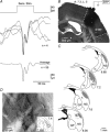
Figure 1
Figure 1. Anatomo-functional characterization of CS projections from barrel cortex
A, average surface ERP (bottom record, n = 55 successive trials), and examples of 4 individual records (middle), in response to sensory stimulations (Sens. Stim., air-puffs of 40 p.s.i. applied to the contralateral whiskers) (top trace). B, microphotograph of the PHA-L injection site in the barrel cortex (dashed lines, S1BF) at the indicated anterior and lateral positions, and from the cortical field where the ERPs shown in A were recorded. C, reconstructions of selected coronal sections (at the indicated anterior position) showing the patterns of CS axonal projections (grey areas) within the ipsilateral dorsolateral striatum, which are concordant with those previously described (see Alloway et al. 1999; Wright et al. 1999). D, microphotograph of PHA-L-labelled CS axons, in the striatal sector shown in the inset, exhibiting the classical densely packed clusters of terminals (see Alloway et al. 1999). cc, corpus callosum; GP, globus pallidus; LV, lateral ventricle; ic, internal capsule; NS, neostriatum; S1BF, barrel field region of primary somatosensory cortex.
Figure 7
Figure 7. Contribution of Cl-dependent synaptic inhibition to sensory integration in MSNs
A, synthetic projection micrograph (from a 120 μm-thick whole mount) of a striatal MSN recorded with a KCl electrode and intracellularly labelled with Neurobiotin. B, barrel cortex ECoG (upper trace) and simultaneously recorded intracellular activity of a Cl-loaded MSN (bottom trace). Note the depolarized membrane potential and the elevated firing rate compared to MSN recorded with a KAc electrode (see Fig. 4C). As shown by the expanded record (inset, calibrations: 10 mV, 10 ms), the spontaneous firing is caused by large-amplitude and long-duration synaptic potentials, probably resulting from additional Cl-dependent synaptic depolarizations. C, superimposition of 3 successive suprathreshold (Ca, the firing probability is indicated) or subthreshold synaptic responses (Cb) evoked in two different Cl-filled MSNs by the same sensory stimulus (top, 40 p.s.i.). The bottom trace in Cb represents the average of 37 successive trials. D, comparison of the percentage of MSNs (and corresponding number of cells) without sensory responses (Non Resp.), exhibiting subthreshold (dPSP Resp.) or suprathreshold responses (AP Resp.) when recorded with KAc or KCl electrodes. E, activation of presumed striatal GABAergic interneurons by whisker deflection. Ea, microphotograph of a putative GABAergic striatal interneuron labelled by juxtacellular injection of Neurobiotin. This cell exhibited the morphological features (for a detailed description, see Results) of striatal interneurons with expanded dendritic field (see Kawaguchi, 1993). The inset shows the short-duration (0.58 ms) extracellular spike of the labelled neuron. Eb, four successive extracellular sensory responses of a putative GABAergic interneuron and corresponding post-stimulus time histogram of action potential discharges (50 collected sweeps, bin = 2 ms). The vertical dashed line indicates the onset of the air-puffs (top).
Figure 4
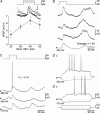
Figure 4. Morpho-functional properties of MSNs located in the striatal projection field of barrel cortex
A, synthetic projection micrograph (from an 80 μm-thick whole mount) of a striatal MSN labelled by intracellular injection of Neurobiotin. This cell exhibited the characteristic morphological features of MSNs (for a detailed description, see Results). B, membrane excitability of barrel cortex-related striatal MSNs. Ba, voltage changes and firing patterns (top records) in a MSN in response to negative and positive current injections (bottom traces). Note the high membrane polarization at rest and the slow ramp depolarization (dashed line) induced by the threshold current pulse, which led to a delayed firing. Increasing intensity of injected current resulted in a reduction of first spike latency and an augmentation of firing rate (grey record). Bb, average voltage changes (ΔV, filled circles) and mean firing rate (F (Hz), open circles) plotted as a function of current pulses of increasing intensity. Each data point corresponds to the mean values calculated from 20 successive trials. The F–I relation was best fitted by a sigmoid curve (r2 = 0.99) and the V–I relation showed a pronounced inward rectification for current pulses more negative than −0.4 nA. C, barrel cortex ECoG (upper trace) and simultaneously recorded intracellular activity of a MSN at rest (−76 mV). Both signals are rhythmic and temporally correlated as evidenced by their oscillatory (∼7 Hz) cross-correlation (inset, scale bar: 200 ms). Note the attenuation of spontaneous intracellular voltage fluctuations after injection of a continuous negative current (−1 nA; −100 mV), which is consistent with the membrane rectification expressed by MSNs (see Bb).
Figure 2
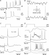
Figure 2. Sensory responses in layer 5 barrel cortex pyramidal cells
A, synthetic projection micrograph (from a 160 μm-thick whole mount) of a Neurobiotin-injected cortical pyramidal neuron whose soma was located in layer 5 of the barrel cortex. Calibration: 50 μm. B, electrophysiological properties of layer 5 neurons. Ba, intracellular responses (top records) to 200 ms positive and negative current pulses (bottom traces) from a neuron exhibiting a regular spiking pattern (top) and another cell generating intrinsic bursting (oblique arrow, bottom). In both cells, the averaging (n = 20) of current-induced hyperpolarizations revealed a sag potential and post-anodal rebound of excitation (oblique crossed arrows). Bb, plots of averaged (n = 20 trials for each intensity) voltage changes (ΔV, filled circles) and mean firing frequency (F (Hz), open circles) as a function of current intensity. Whereas the V–I relation, measured at the time indicated by the open circle in Ba, was linear (linear fit; r2 = 0.99), the F–I relation followed a sigmoid function (r2 = 0.99). C, sensory responses induced in layer 5 barrel cortex cells by contralateral whisker deflection. Ca, sensory stimuli (top traces), applied with increasing air-puff pressures, induced depolarizing postsynaptic potentials (dPSPs) that could eventually reach the firing threshold (bottom records). As depicted in the graph below, both dPSPs and firing probability increased in parallel with the intensity of the sensory stimulus (n = 50 trials for each intensity). Cb and c, typical examples of layer 5 cells responding (3 successive individual responses) to air-puffs of same intensity (top traces) with dPSPs that could fired action potentials (Cb) or remain subthreshold (Cc). In Cb, the firing probability (PF) calculated from 50 trials is indicated and, in Cc, the bottom trace represents the average response to 36 stimuli. Here and in the following figures, the values of membrane potential are indicated to the left of the intracellular records.
Figure 3
Figure 3. Responses of identified barrel cortex CS neurons to whisker stimulation
A, antidromic identification of CS neurons. Superimposition of 3 successive responses of a barrel cortex layer 5 neuron to electrical stimulations (vertical arrow) of the ipsilateral striatum (top traces). Note the short (0.64 ms), and stable, latency of the first evoked action potential, which was abolished (bottom record) by the collision with a spontaneously occurring orthodromic spike (oblique arrow). This cell displayed an intrinsic bursting pattern (inset) in response to positive current (+0.6 nA) injection (calibrations: 20 mV, 50 ms). B, continuous (1.4 s) intracellular recording (bottom record) from an identified CS neuron and the corresponding surface ECoG activity (top trace). The inset depicts the cross-correlation between both signals, using the cortical surface activity as reference (calibration, 200 ms). Note the high correlation between the two oscillatory signals, at a frequency of ∼7.5 Hz, with a temporal shift of −18.7 ms. C, three successive individual responses in a CS neuron (same as in B) to air-puffs applied on the contralateral whiskers (top). Note the high firing probability (PF) of the cell in response to sensory stimuli despite the occurrence of a small subthreshold dPSP (asterisk), apparently shunted by a prior synaptic activity. D, barrel cortex ERPs (top records) and corresponding intracellular responses (n = 2) recorded from an identified CS cell (black traces) and an ‘unidentified’ layer 5 cell (grey traces) in the same experiment and in response to the same sensory stimulus (upper trace). As shown by the expanded records (top inset, calibrations: 10 mV, 10 ms), the latency and shape of suprathreshold sensory responses (spikes are truncated) in both cells were very similar. Injection of a positive current pulse (0.6 nA) generated an intrinsic bursting firing pattern in the two neurons (bottom inset, calibrations: 20 mV, 50 ms).
Figure 5
Figure 5. Sensory responses in striatal MSNs
A, air-puffs applied to contralateral whiskers, with increasing pressures (top traces) evoked in the recorded MSN subthreshold dPSPs of variable amplitude (bottom records, calibrations: 5 mV, 10 ms). The graph below depicts the mean amplitude of dPSPs (n > 22 trials for each intensity) as a function of the intensity of sensory stimuli (Sens. Stim.). Note the inflection in the amplitude of the sensory response above 40 p.s.i., which corresponded to the stimulus producing the largest response in the corresponding cortical ERP (‘optimal stimulus’, see Methods). B, examples of dPSPs (n = 3) and corresponding average potential (n = 40 successive trials) evoked in MSNs by a whisker stimulus of 40 p.s.i. (top trace). In this cell, the sensory responses remained subthreshold. C, three successive responses in a MSN that could respond to the optimal sensory stimulus (top), by sub- or suprathreshold dPSPs with the indicated firing probability (PF). D, current-induced (bottom) responses recorded from the MSNs shown in B (Da) and in C (Db).
Figure 6
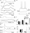
Figure 6. The variability of sensory responses in striatal MSNs is not due to distinct membrane excitability
A, electrophysiological properties of MSNs that did not respond to sensory stimuli. Aa, four successive sweeps recorded in a MSN during application of sensory stimuli of 40 p.s.i. Although some synaptic depolarizations occurred in a temporal window compatible with sensory-evoked events (see Fig. 5B and C), the averaging of 50 successive trials revealed a complete lack of sensory responses (bottom trace). As indicated by the large-amplitude ERP (top average record), the absence of whisker-induced responses in the striatal cell did not result from a deprivation of sensory responsiveness in barrel cortex neurons. Ab, current-induced (bottom traces) voltage responses (top) in the MSN shown in Aa. Note the similarity of cell excitability compared to that of sensory-reactive MSNs (see Fig. 5Da and Db). B, the same sensory stimulus (top trace) can generate, in two neighbouring MSNs, either no response (Ba, two individual records and corresponding average sweep) or dPSPs that can cause spike discharge (Bb, three individual records), with the indicated probability firing (PF). In Bb, spikes are truncated for convenience. C, pooled histograms of membrane potential (Vm, mV), input resistance (Rm, MΩ), membrane time constant (τm, ms), action potential threshold (APth, mV) and mean spontaneous firing rate (SFR, Hz) calculated from MSNs without sensory responses (Non resp., n = 22 neurons) and exhibiting sub- (dPSP resp., n = 19 neurons) or suprathreshold responses (AP resp., n = 8 neurons). None of these parameters was found to be significantly different between the three neuronal groups (P > 0.05 for each parameter).
Figure 8
Figure 8. Comparison of sensory-mediated responses in barrel cortex layer 5 cells and MSNs
A, comparison of the percentage of MSNs and barrel cortex layer 5 neurons (Cx), and corresponding number of recorded cells, exhibiting no detectable sensory response (Non Resp.), subthreshold (dPSP Resp.) or suprathreshold responses (AP Resp.). B, summary histograms of probability of firing (PF), latency of firing (LF) and corresponding values of standard deviation (σLF), calculated from MSNs (n = 8) and cortical neurons (n = 58) that could be fired by the sensory stimulus. Only the probability of firing was significantly different between the two groups (P = 0.03). C, comparison of sensory responses evoked in layer 5 cortical cells and MSNs recorded during the same experiment. Ca, averaged ERPs (top records) and superimposition (n = 3) of corresponding intracellular activities recorded, during the same experiment and following the same whisker deflection, from a layer 5 barrel cortex neuron responding by suprathreshold responses (Cx AP Resp., PF = 0.74) (spikes are truncated), a MSN responding by subthreshold depolarizations (MSN dPSP Resp.) and another MSN without detectable response (MSN Non Resp.). Cb, synthetic representation of sensory responses in barrel cortex layer 5 neurons (Cx) and MSNs that were recorded during the same experiment and after application of identical whisker stimulations (n = 17 experiments). Each vertical dashed line corresponds to a single experiment and the corresponding values of dPSP amplitude, probability of firing, as well as the number of unresponsive neurons (Non Resp.) are indicated. The vertical arrow indicates the experiment illustrated in Ca.

Similar articles

Cited by

References

    1. Alloway KD, Crist J, Mutic JJ, Roy SA. Corticostriatal projections from rat barrel cortex have an anisotropic organization that correlates with vibrissal whisking behavior. J Neurosci. 1999;19:10908–10922. - PMC - PubMed
    1. Alloway KD, Lou L, Nwabueze-Ogbo F, Chakrabarti S. Topography of cortical projections to the dorsolateral neostriatum in rats: multiple overlapping sensorimotor pathways. J Comp Neurol. 2006;499:33–48. - PubMed
    1. Azouz R, Gray CM. Cellular mechanisms contributing to response variability of cortical neurons in vivo. J Neurosci. 1999;19:2209–2223. - PMC - PubMed
    1. Barnes TD, Kubota Y, Hu D, Jin DZ, Graybiel AM. Activity of striatal neurons reflects dynamic encoding and recoding of procedural memories. Nature. 2005;437:1158–1161. - PubMed
    1. Brecht M. Barrel cortex and whisker-mediated behaviors. Curr Opin Neurobiol. 2007;17:408–416. - PubMed

Publication types

LinkOut - more resources