. 2010 Nov 15;191(4):731-9.
doi: 10.1083/jcb.201007107. Epub 2010 Nov 8.

Cep152 acts as a scaffold for recruitment of Plk4 and CPAP to the centrosome

Affiliations

Cep152 acts as a scaffold for recruitment of Plk4 and CPAP to the centrosome

Onur Cizmecioglu et al. J Cell Biol. .

Abstract

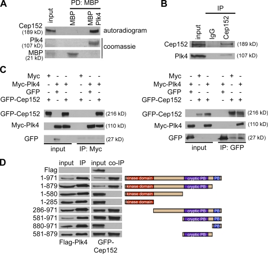
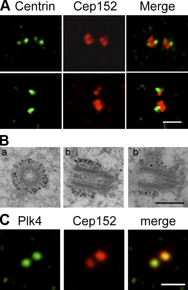
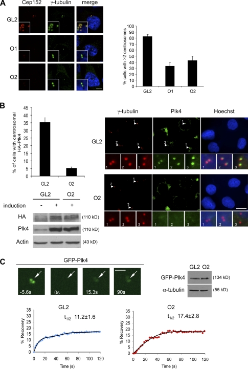
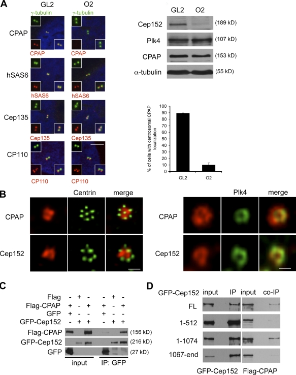
Both gain and loss of function studies have identified the Polo-like kinase Plk4/Sak as a crucial regulator of centriole biogenesis, but the mechanisms governing centrosome duplication are incompletely understood. In this study, we show that the pericentriolar material protein, Cep152, interacts with the distinctive cryptic Polo-box of Plk4 via its N-terminal domain and is required for Plk4-induced centriole overduplication. Reduction of endogenous Cep152 levels results in a failure in centriole duplication, loss of centrioles, and formation of monopolar mitotic spindles. Interfering with Cep152 function prevents recruitment of Plk4 to the centrosome and promotes loss of CPAP, a protein required for the control of centriole length in Plk4-regulated centriole biogenesis. Our results suggest that Cep152 recruits Plk4 and CPAP to the centrosome to ensure a faithful centrosome duplication process.


Figures

Figure 1.
Figure 1.
Cep152 interacts with Plk4 in vitro and in vivo. (A) To analyze in vitro binding between Cep152 and Plk4, either MBP tag alone or MBP-tagged recombinant Plk4 immobilized on amylose beads was used in a binding assay with in vitro–translated [35S]Cep152. Binding of [35S]Cep152 to Plk4 was detected by autoradiography. Equal pull-down of MBP and MBP-Plk4 was shown by Coomassie staining. PD, pull-down. (B) Endogenous Cep152 was immunoprecipitated from U2OS cell extracts using Cep152 (Ab1140). Coprecipitated endogenous Plk4 was detected with a mouse anti-Plk4 antibody by Western blotting. Immunoprecipitation control, random rabbit IgGs. (C) Coimmunoprecipitation of Myc-Plk4 and GFP-Cep152 after coexpression in 293T cells. Reciprocal immunoprecipitation Western blots were performed with anti-Myc and anti-GFP antibodies. Coprecipitated proteins were detected by Western blotting against the corresponding tag. (D, left) Different Flag-Plk4 fragments were coexpressed with GFP-Cep152 in 293T cells. Anti-Flag immunoprecipitates were analyzed in immunoblots for coprecipitated GFP-Cep152 using GFP antibodies. (right) Scheme of expressed Plk4 fragments.
Figure 2.
Figure 2.
Cep152 localizes to the PCM. (A) Immunofluorescence images showing that a minor fraction of endogenous Cep152 (red) colocalizes with the centriolar marker centrin-2 (green) in U2OS cells. (top) Cell with four centrioles. (bottom) Cell with two centrioles. (B) Immunogold EM of U2OS cells shows that Cep152 localizes to the PCM cloud surrounding the outer wall at the proximal ends of the centriole. Cep152 localization on transversial sections (a) and longitudinal sections of one centriole (b and b’) are shown. (C) Costaining of U2OS cells with antibodies against Cep152 (red) and Plk4 (green). Bars: (A and C) 2 µm; (B) 0.5 µm.
Figure 3.
Figure 3.
Cep152 is required for centriole duplication. (A) U2OS cells were transfected with siRNAs against Cdk2 (positive control), Cep152 (O1 and O2), GL2 (negative control), and arrested in S phase by aphidicolin treatment. 70 h later, cells with more than four centrioles were counted. Red, Cep152; green, centrin-2; blue, DNA. (B) U2OS cells were transfected with either GL2 or Cep152 siRNAs (O1 or O2). Spindle poles were depicted with centrin staining (green, centrin-2) and mitotic spindles with α-tubulin antibodies (red) or DNA (blue). 72 h after transfection, cells with monopolar or bipolar mitotic spindles were counted. (C) U2OS cells were transfected with either GFP or GFP-Cep152 1–512 (green). Centrioles were visualized with centrin-2 staining (red). 48 h and 72 h after transfection, cells with less than two centrioles were counted. (left) Representative pictures of the observed phenotypes. (D) Cep152 full length (FL), Cep152 fragments (1–512 and 508–end), or GFP were overexpressed in U2OS cells. 48 h after transfection, cells with or without centriolar Plk4 staining (blue) were counted. GT335 (red; Bobinnec et al., 1998), an antibody to modified tubulin, was used as a marker for centrosomes. Insets show enlargements of centrosomes as merged image and individual channels. Error bars show the SDs of at least three independent experiments. Bars, 5 µm.
Figure 4.
Figure 4.
Cep152 recruits Plk4 to the centrosome. (A) Centriole overduplication in HeLa Tet-on cells was forced by induction of Plk4 expression through addition of doxycycline. Simultaneously, cells were transfected with either GL2 or one of two different Cep152 siRNAs (O1 or O2). Centrosomes were stained with γ-tubulin (green) antibodies or Cep152 (red). 72 h after transfection, cells with less than two centrosomes were counted. (B) Hela Tet-on cells were transfected with GL2 or Cep152 siRNAs (O1 and O2) treated with aphidicolin for 24 h. Plk4 expression was induced in the last 20 h of aphidicolin treatment. Cells were scored for centrosomal HA-Plk4 signal. Induced HA-Plk4 signal (green) locates to the centrosome (red, γ-tubulin). Blue, DNA. (right) Insets display enlargements of the selected regions in the indicated channels (arrowheads). The last set of insets includes a triple merge. Corresponding immunoblots from siRNA-treated and induced cells were analyzed for Plk4 levels using antibodies against HA and Plk4 in comparison with uninduced, control siRNA-treated samples. (A and B) Error bars indicate SDs (n = 3). (C) Dynamics of Plk4 at centrosomes in response to Cep152 RNAi. FRAP was performed on U2OS cells treated with either GL2 or Cep152 siRNAs (O2) for 60 h followed by GFP-Plk4 transfections for 14 h. GFP-Plk4–positive, unsplit centrosomes in the same plane of focus were selected for photobleaching and subsequent imaging. 80 × 80–pixel squares surrounding the centrosome were bleached (bleach time 2.5 s), and the recovery of GFP fluorescence on centrosomes was imaged over time. (left) Arrows mark photobleached regions on the centrosome. (right) Relative expression levels of GFP-Plk4 in GL2- and O2-transfected cells were determined. (bottom) Mean fluorescence recovery profiles of GFP-Plk4 on the centrosome were depicted in GL2- and O2-treated cells (n = 12). Mean t1/2 of experiments is shown ± SD. Student’s t test was performed with GL2 or Cep152 siRNA-treated cells for recovery of GFP-Plk4 on the centrosome with P < 0.0001. Bars: (A) 5 µm; (B) 20 µm; (C) 3 µm.
Figure 5.
Figure 5.
CPAP localization at the centrosome is dependent on Cep152. (A) U2OS cells were transfected with either GL2 or Cep152 siRNAs. Colocalizations of CPAP, hSas6, Cep135, and CP110 (red) together with γ-tubulin (green) were determined by immunofluorescence. Insets show enlargements of the merged image and individual channels. (right) Protein levels of the indicated proteins were determined by Western blotting. The graph shows a quantification of the percentage of cells with centrosomal CPAP localization. Error bars indicate SDs (n = 3). (B, left) Cep152 and CPAP costainings (red) within the flower-like centrin-2 structures (green) were depicted. (B, right) Cep152 and CPAP colocalizations (red) were performed together with HA-Plk4 (green) using anti-HA antibodies. (C) Flag-CPAP and GFP or GFP-Cep152 constructs were coexpressed in 293T cells. GFP and GFP-Cep152 were immunoprecipitated 48 h after expression. Coimmunoprecipitated Flag-CPAP was detected by Western blotting against the Flag tag. (D) Different GFP-Cep152 fragments (Fig. S3 A) were coexpressed with Flag-CPAP in 293T cells. Anti-GFP immunoprecipitates were analyzed by Western blotting for coprecipitated Flag-CPAP with anti-Flag antibodies. Bars: (A) 5 µm; (B, left) 1 µm; (B, right) 0.5 µm.

Similar articles

Cited by

References

    1. Andersen J.S., Wilkinson C.J., Mayor T., Mortensen P., Nigg E.A., Mann M. 2003. Proteomic characterization of the human centrosome by protein correlation profiling. Nature. 426:570–574 10.1038/nature02166 - DOI - PubMed
    1. Basto R., Brunk K., Vinadogrova T., Peel N., Franz A., Khodjakov A., Raff J.W. 2008. Centrosome amplification can initiate tumorigenesis in flies. Cell. 133:1032–1042 10.1016/j.cell.2008.05.039 - DOI - PMC - PubMed
    1. Bettencourt-Dias M., Glover D.M. 2007. Centrosome biogenesis and function: centrosomics brings new understanding. Nat. Rev. Mol. Cell Biol. 8:451–463 10.1038/nrm2180 - DOI - PubMed
    1. Bettencourt-Dias M., Rodrigues-Martins A., Carpenter L., Riparbelli M., Lehmann L., Gatt M.K., Carmo N., Balloux F., Callaini G., Glover D.M. 2005. SAK/PLK4 is required for centriole duplication and flagella development. Curr. Biol. 15:2199–2207 10.1016/j.cub.2005.11.042 - DOI - PubMed
    1. Blachon S., Gopalakrishnan J., Omori Y., Polyanovsky A., Church A., Nicastro D., Malicki J., Avidor-Reiss T. 2008. Drosophila asterless and vertebrate Cep152 Are orthologs essential for centriole duplication. Genetics. 180:2081–2094 10.1534/genetics.108.095141 - DOI - PMC - PubMed

Publication types

MeSH terms