. 2010 Nov 15;191(4):721-9.
doi: 10.1083/jcb.201006049. Epub 2010 Nov 8.

Cep152 interacts with Plk4 and is required for centriole duplication

Affiliations

Cep152 interacts with Plk4 and is required for centriole duplication

Emily M Hatch et al. J Cell Biol. .

Abstract

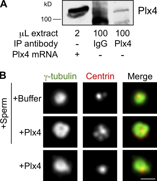
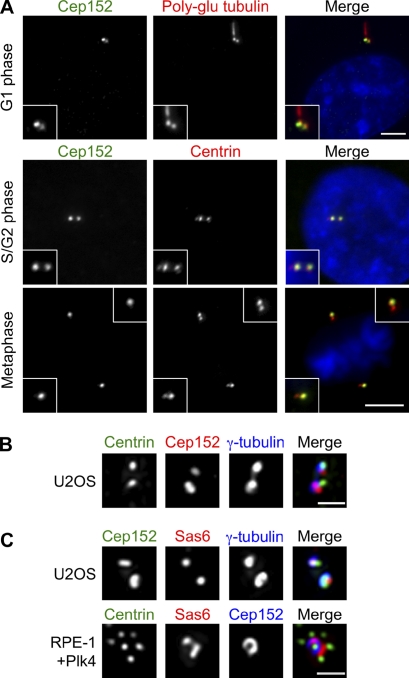
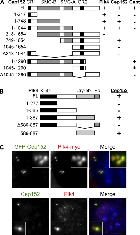
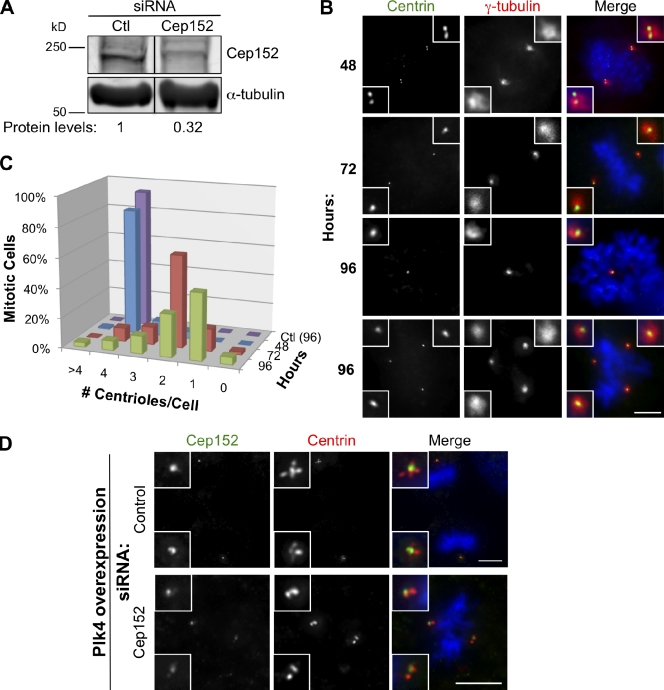
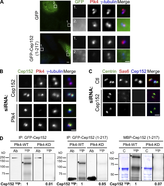
Centrioles are microtubule-based structures that organize the centrosome and nucleate cilia. Centrioles duplicate once per cell cycle, and duplication requires Plk4, a member of the Polo-like kinase family; however, the mechanism linking Plk4 activity and centriole formation is unknown. In this study, we show in human and frog cells that Plk4 interacts with the centrosome protein Cep152, the orthologue of Drosophila melanogaster Asterless. The interaction requires the N-terminal 217 residues of Cep152 and the crypto Polo-box of Plk4. Cep152 and Plk4 colocalize at the centriole throughout the cell cycle. Overexpression of Cep152 (1-217) mislocalizes Plk4, but both Cep152 and Plk4 are able to localize to the centriole independently of the other. Depletion of Cep152 prevents both normal centriole duplication and Plk4-induced centriole amplification and results in a failure to localize Sas6 to the centriole, an early step in duplication. Cep152 can be phosphorylated by Plk4 in vitro, suggesting that Cep152 acts with Plk4 to initiate centriole formation.


Figures

Figure 1.
Figure 1.
Plx4 overexpression drives centriole amplification in Xenopus egg extract. (A) Western blot of Xenopus egg extracts after addition of Plx4 mRNA or immunoprecipitation (IP) of endogenous Plx4 using anti-Plx4 or nonimmune IgG as a control. Immunoblotted with anti-Plx4. (B) Xenopus egg extract incubated with Plx4 or no mRNA with or without Xenopus sperm. Centrin and γ-tubulin mark the centrioles and pericentriolar material, respectively. Bar, 1 µm.
Figure 2.
Figure 2.
Cep152 localizes to centrioles throughout the cell cycle. (A) RPE-1 cells at the indicated cell cycle stages were fixed and stained for Cep152, centrin, or polyglutamylated (poly-glu) tubulin, which marks centrioles and primary cilia, and DNA. Insets show enlarged centrosomes. Bars, 5 µm. (B) G1 centrosomes in a U2OS cell stained for Cep152, centrin, and γ-tubulin. (C, top) Centrosomes in a U2OS cell stained for Cep152, Sas6, and γ-tubulin. (bottom) Centrosome in a RPE-1 cell overexpressing Plk4 stained for centrin, Sas6, and Cep152. (B and C) Images are maximum projections of deconvoluted image stacks. Bars, 1 µm.
Figure 3.
Figure 3.
Cep152 and Plk4 interaction and localization. (A) Schematic of Cep152 full length (FL) and deletion constructs showing the ability to interact with Plk4 and Cep152 and to localize to the centrosome (Cent). Interactions were determined by coimmunoprecipitation of GFP-Cep152 constructs from HEK293T lysates with Plk4-myc or Cep152-myc. Centrosome localization was tested by expression of fragments as GFP fusions in U2OS cells. Numbers indicate Cep152 amino acids. CR1 and CR2 indicate conserved domains in Cep152 orthologues; SMC-A and SMC-B indicate coiled-coil regions. (B) Schematic of Plk4 deletion constructs and summary of interactions with Cep152. Domains: Cry-pb, crypto Polo-box; Pb, Polo-box; KinD, kinase domain. Interactions were determined by coimmunoprecipitation of Plk4-myc fragments from HEK293T lysates with GFP-Cep152. (C, top) U2OS cells expressing GFP-Cep152 and Plk4-myc labeled with antibodies to GFP and myc. (bottom) Untransfected U2OS cells labeled with antibodies to Cep152 and Plk4. Insets show enlarged centrosomes. Bar, 5 µm.
Figure 4.
Figure 4.
Cep152 is required for centriole duplication and amplification. (A) Western blotting of U2OS cell lysates 72 h after transfection with control (Ctl) siRNAs or Cep152 siRNAs immunoblotted for Cep152 or α-tubulin as a loading control. The indicated Cep152 protein levels are normalized to α-tubulin levels. Black lines indicate that intervening lanes have been spliced out. (B) U2OS cells were fixed at the indicated times after Cep152 siRNA transfection, and centriole number was determined by labeling for centrin and γ-tubulin. Insets show enlarged centrosomes. (C) Quantification of B. Centriole number in cells transfected with control siRNAs for 96 h was determined as described above. n = 200 cells over three experiments for each time point. (D) RPE-1 cells bearing Tet-inducible Plk4 were depleted of Cep152 by siRNA for 72 h then treated with doxycycline for 18 h to induce Plk4 expression. Centriole number was determined by labeling for centrin, and depleted cells were identified by reduced Cep152 staining. (B and D) Insets show enlarged centrosomes. Bars, 5 µm.
Figure 5.
Figure 5.
Functional interaction of Cep152 and Plk4. (A) Plk4 is lost from the centrosome in cells expressing GFP-Cep152 (1–217). U2OS cells transfected with GFP or GFP-Cep152 (1–217) were fixed 24 h after transfection and labeled with antibodies to GFP, Plk4, and γ-tubulin. Numbered boxes identify enlarged images shown on right. Bars (boxes) 1 µm; (fields) 5 µm. (B) U2OS cells were depleted of Plk4 or Cep152 by siRNA transfection, fixed 72 h after transfection, and labeled with antibodies to Cep152, Plk4, and γ-tubulin. Image pairs represent centrosomes in cells from the same coverslip taken with the same camera settings. (C) U2OS cells transfected for 72 h with either control siRNA (Ctl) or Cep152 siRNAs were arrested in S phase by incubation in aphidicolin for 24 h and fixed and labeled with antibodies to centrin, Sas6, and Cep152. 25% of Cep152-depleted cells had reduced Sas6 labeling on both centrioles (middle), 65% had no Sas6 labeling (bottom), and 10% had centrioles that differed in the amount of Sas6 (n = 150 cells). Exposure and gain conditions were constant for Sas6 image acquisition. (B and C) Bars, 1 µm. (D) Cep152 can be phosphorylated by Plk4 in vitro. Purified GST-Plk4, either wild type (WT) or kinase dead (KD), was incubated in the presence of [32P]ATP (32P) with full-length GFP-Cep152 (left) or GFP-Cep152 (1–217; middle) isolated from HEK293T cells by anti-GFP immunoprecipitation (IP) or with MBP-Cep152 (1–217; right) purified from E. coli. Proteins were visualized by Western blotting (Ab) of 0.1× kinase assay input using antibodies to GFP (left and middle) or by Coomassie stain (C) of the gel (right). Bands marked with an asterisk indicate autophosphorylated Plk4. Cep152 32P incorporation was normalized within each gel group. Black lines indicate that gel lanes imaged by different methods were spliced together.

References

    1. Andersen J.S., Wilkinson C.J., Mayor T., Mortensen P., Nigg E.A., Mann M. 2003. Proteomic characterization of the human centrosome by protein correlation profiling. Nature. 426:570–574 10.1038/nature02166 - DOI - PubMed
    1. Basto R., Lau J., Vinogradova T., Gardiol A., Woods C.G., Khodjakov A., Raff J.W. 2006. Flies without centrioles. Cell. 125:1375–1386 10.1016/j.cell.2006.05.025 - DOI - PubMed
    1. Basto R., Brunk K., Vinadogrova T., Peel N., Franz A., Khodjakov A., Raff J.W. 2008. Centrosome amplification can initiate tumorigenesis in flies. Cell. 133:1032–1042 10.1016/j.cell.2008.05.039 - DOI - PMC - PubMed
    1. Blachon S., Gopalakrishnan J., Omori Y., Polyanovsky A., Church A., Nicastro D., Malicki J., Avidor-Reiss T. 2008. Drosophila asterless and vertebrate Cep152 Are orthologs essential for centriole duplication. Genetics. 180:2081–2094 10.1534/genetics.108.095141 - DOI - PMC - PubMed
    1. Carvalho-Santos Z., Machado P., Branco P., Tavares-Cadete F., Rodrigues-Martins A., Pereira-Leal J.B., Bettencourt-Dias M. 2010. Stepwise evolution of the centriole-assembly pathway. J. Cell Sci. 123:1414–1426 10.1242/jcs.064931 - DOI - PubMed

Publication types