. 2010 Nov 23;107(47):20370-5.
doi: 10.1073/pnas.1014194107. Epub 2010 Nov 8.

Human tumor virus utilizes exosomes for intercellular communication

Affiliations

Human tumor virus utilizes exosomes for intercellular communication

David G Meckes Jr et al. Proc Natl Acad Sci U S A. .

Abstract

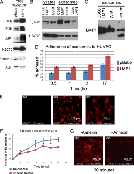
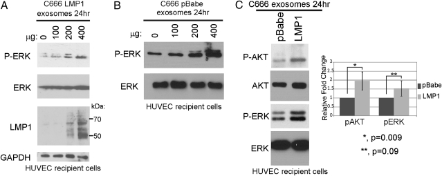
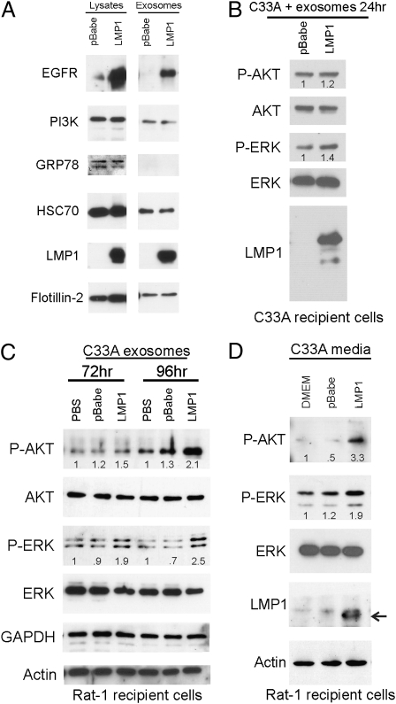
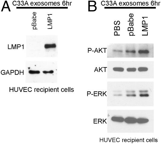
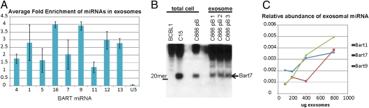
The Epstein-Barr virus (EBV) latent membrane protein 1 (LMP1) is expressed in multiple human malignancies and has potent effects on cell growth. It has been detected in exosomes and shown to inhibit immune function. Exosomes are small secreted cellular vesicles that contain proteins, mRNAs, and microRNAs (miRNAs). When produced by malignant cells, they can promote angiogenesis, cell proliferation, tumor-cell invasion, and immune evasion. In this study, exosomes released from nasopharyngeal carcinoma (NPC) cells harboring latent EBV were shown to contain LMP1, signal transduction molecules, and virus-encoded miRNAs. Exposure to these NPC exosomes activated the ERK and AKT signaling pathways in the recipient cells. Interestingly, NPC exosomes also contained viral miRNAs, several of which were enriched in comparison with their intracellular levels. LMP1 induces expression of the EGF receptor in an EBV-negative epithelial cell line, and exosomes produced by these cells also contain high levels of EGF receptor in exosomes. These findings suggest that the effects of EBV and LMP1 on cellular expression also modulate exosome content and properties. The exosomes may manipulate the tumor microenvironment to influence the growth of neighboring cells through the intercellular transfer of LMP1, signaling molecules, and viral miRNAs.


Conflict of interest statement

The authors declare no conflict of interest.

Figures

Fig. 1.
Fig. 1.
Characterization of NPC exosomes and their uptake by HUVECs. (A) Purified exosomes from conditioned media of C666-pBabe and C666-LMP1 were analyzed by immunoblotting with the indicated antibodies and the LMP1 S12 monoclonal antibody. (B) Levels of LMP1 within cellular lysates were compared between C666-LMP1 and B958 cells and in exosomes produced by C666-LMP1, B958, and C33A-LMP1. (C) Exosomes from cultured C666 cells, C15 xenograft, or C15 mouse serum were analyzed by immunoblotting for LMP1 levels by using pooled LMP1 rat monoclonal antibodies. (D and E) Three separate preparations of purified exosomes were labeled with DiI and incubated with HUVECs for the indicated times. Fluorescence was monitored in a plate reader (D) or by confocal microscopy (E). The numbers were normalized to equal DiI totals and represented as a percentage of exosomes adhered. (F and G) Annexin blocking of exosome fusion was measured by fluorescence dequenching of R18-labeled exosomes at the indicated times by using a plate reader (F) or visualized by confocal microscopy (G).
Fig. 2.
Fig. 2.
Transfer of LMP1 and activation of ERK and AKT pathways in HUVECs by NPC exosomes. HUVECs were incubated with increasing amounts of purified C666-LMP1 (A) or C666-pBabe (B) exosomes for 24 h in serum-free media. Cell lysates were analyzed by immunoblotting for the indicated proteins and S12 mAb for LMP1. (C) HUVECs exposed to 400 μg of pBabe or LMP1 exosomes for 24 h were analyzed by immunoblotting for activated ERK and AKT. Levels of pAKT and pERK normalized to total AKT and ERK protein levels and represented relative to pBabe exosomes are shown from three independent experiments.
Fig. 3.
Fig. 3.
Exosomes and conditioned media from LMP1-expressing C33A cells activate ERK and AKT in recipient cells. (A) Cell lysates and purified exosomes from C33A-LMP1 or C33A-pBabe cells were analyzed by immunoblotting with antibodies against EGFR, p85, GRP78, HSC70, LMP1, flotillin-2, actin, and S12 mAb for LMP1. (B) Untransfected C33A cells were incubated with C33A-pBabe or LMP1 exosomes for 24 h in serum-free media, and the indicated proteins were identified by immunoblotting of cell lysates. (C) C33A exosomes were incubated with Rat-1 cells for 72 or 96 h, and cell lysates were analyzed for the indicated proteins by immunoblotting. Numbers indicate fold change over PBS control. (D) Media alone (DMEM) or conditioned media from C33A-pBabe or C33A-LMP1 was clarified and added to Rat-1 cells every 24 h for 5 d. pAKT and pERK intensities in all experiments were normalized to total protein levels, and the relative values to the control are indicated in each channel. Representative blots from two to four independent experiments are shown.
Fig. 4.
Fig. 4.
Activation of ERK and AKT pathways in HUVECs exposed to C33A-LMP1 exosomes. HUVECs were incubated for 6 h with PBS, pBabe, and LMP1 exosomes, and recipient cell lysates were immunoblotted for LMP1 with the S12 monoclonal antibody (A) or pAKT and pERK (B) with AKT and ERK as loading controls. Representative results from three independent experiments are shown.
Fig. 5.
Fig. 5.
Viral miRNAs are enriched in NPC exosomes and taken up by HUVECs. (A) Total RNA from C666 cells or exosomes was analyzed by using quantitative RT-PCR for the BART miRNAs with the small nuclear RNA U5A as a negative control. Data are represented as relative levels of the individual miRNAs in comparison with their intracellular levels in equal amounts of C666 total cell RNA and exosomal RNA from three independent experiments run in triplicate. (B) Equal amounts of total cell RNA from C666 cells or three independent exosome preps were separated in tris-borate-EDTA gels and transferred to Hybond N+, hybridized with an end-labeled anti-sense oligo to the miRNA-Bart7, and visualized by autoradiography. EBV-negative BCBL1 cells were used as a negative control, and the C15 xenograft was used as a positive control. (C) HUVEC cells were exposed to C666 exosomes for 6 h, and miRNA uptake was monitored by quantitative RT-PCR of total RNA isolated from HUVECs that had been extensively washed to remove unbound exosomes. miRNA levels are represented as relative to the amount of each miRNA present within an equivalent amount of C666 cellular RNA.

References

    1. Raab-Traub N. Epstein–Barr virus in the pathogenesis of NPC. Semin Cancer Biol. 2002;12:431–441. - PubMed
    1. Kaye KM, Izumi KM, Kieff E. Epstein–Barr virus latent membrane protein 1 is essential for B-lymphocyte growth transformation. Proc Natl Acad Sci USA. 1993;90:9150–9154. - PMC - PubMed
    1. Wang D, Liebowitz D, Kieff E. An EBV membrane protein expressed in immortalized lymphocytes transforms established rodent cells. Cell. 1985;43:831–840. - PubMed
    1. Morris MA, Dawson CW, Young LS. Role of the Epstein–Barr virus-encoded latent membrane protein-1, LMP1, in the pathogenesis of nasopharyngeal carcinoma. Future Oncol. 2009;5:811–825. - PubMed
    1. Miller WE, Earp HS, Raab-Traub N. The Epstein–Barr virus latent membrane protein 1 induces expression of the epidermal growth factor receptor. J Virol. 1995;69:4390–4398. - PMC - PubMed

Publication types

MeSH terms

Substances