. 2010 Nov 30;107(48):20744-9.
doi: 10.1073/pnas.1013893107. Epub 2010 Nov 8.

Force-induced formation and propagation of adhesion nanodomains in living fungal cells

Affiliations

Force-induced formation and propagation of adhesion nanodomains in living fungal cells

David Alsteens et al. Proc Natl Acad Sci U S A. .

Abstract

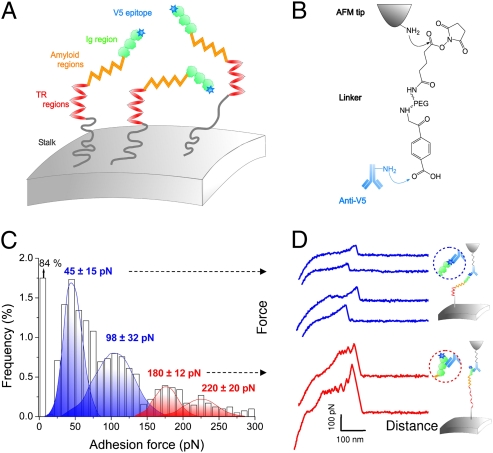
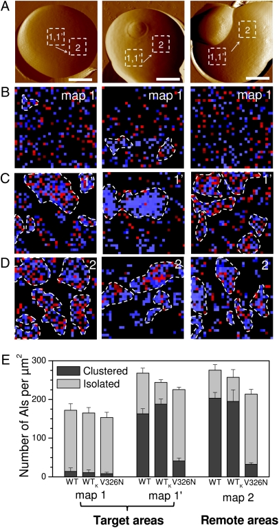
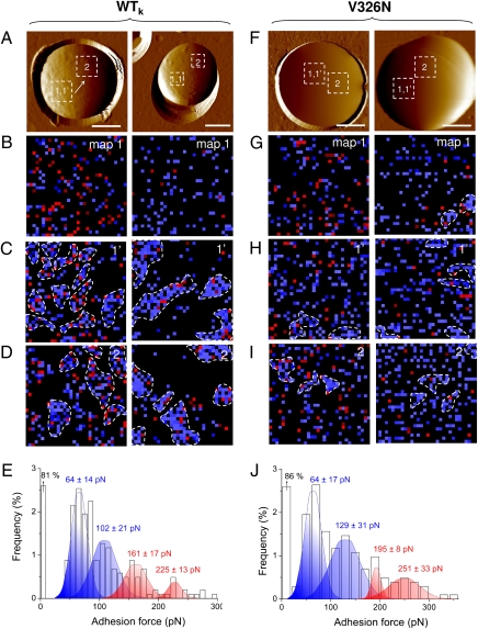
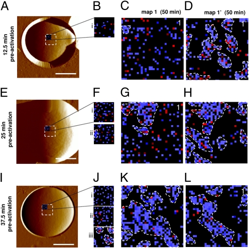
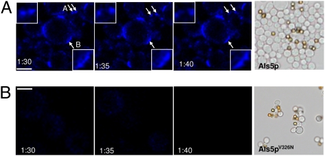
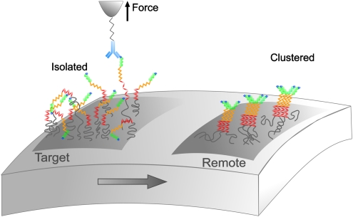
Understanding how cell adhesion proteins form adhesion domains is a key challenge in cell biology. Here, we use single-molecule atomic force microscopy (AFM) to demonstrate the force-induced formation and propagation of adhesion nanodomains in living fungal cells, focusing on the covalently anchored cell-wall protein Als5p from Candida albicans. We show that pulling on single adhesins with AFM tips terminated with specific antibodies triggers the formation of adhesion domains of 100-500 nm and that the force-induced nanodomains propagate over the entire cell surface. Control experiments (with cells lacking Als5p, single-site mutation in the protein, bare tips, and tips modified with irrelevant antibodies) demonstrate that Als5p nanodomains result from protein redistribution triggered by force-induced conformational changes in the initially probed proteins, rather than from nonspecific cell-wall perturbations. Als5p remodeling is independent of cellular metabolic activity because heat-killed cells show the same behavior as live cells. Using AFM and fluorescence microscopy, we also find that nanodomains are formed within ∼30 min and migrate at a speed of ∼20 nm·min(-1), indicating that domain formation and propagation are slow, time-dependent processes. These results demonstrate that mechanical stimuli can trigger adhesion nanodomains in fungal cells and suggest that the force-induced clustering of adhesins may be a mechanism for activating cell adhesion.


Conflict of interest statement

The authors declare no conflict of interest.

Figures

Fig. 1.
Fig. 1.
Detection and unfolding of single Als5p proteins in live cells. (A and B) Principle of the single-molecule force experiment. S. cerevisiae cells expressing Als5p proteins tagged with a V5 epitope (A) are probed, in buffer, using AFM tips terminated with anti-V5 antibodies (B). (C and D) Adhesion force histogram (n = 4,096) from four maps of 1,024 data points (C) and representative force curves (D) obtained by recording spatially resolved force curves over 1 × 1-μm areas of the cell surface using anti-V5 tips. The anti-V5 tip is capable of dual detection: the blue curves show single weak adhesion peaks reflecting recognition of the V5-tag, and the red curves feature sawtooth patterns with multiple force peaks documenting the unfolding of the entire protein via Ig binding. All curves were obtained at 20 °C using a loading rate of 10,000 pN·s−1 and an interaction time of 500 ms. Similar data were obtained using different tips and cells (10 tips, 6 cells). Als proteins expressed on S. cerevisiae exhibit the same activities as they do in C. albicans (13, 14).
Fig. 2.
Fig. 2.
Formation and propagation of Als5p nanodomains. (A) AFM topographic images (scale bars: 1.5 μm), in buffer, showing three different wild-type S. cerevisiae cells expressing V5-tagged Als5p proteins. (B) Adhesion force maps (1 × 1 μm) recorded with an anti-V5 tip on a given target area of the native cells, i.e., cells that were never subjected to force (map 1; recorded on the dashed squares in A). Blue and red pixels correspond to forces smaller and larger than 150 pN, respectively, and thus to V5-tagged Als5p recognition and unfolding. (C) Second adhesion force maps (1 × 1 μm) recorded on the same target area (map 1′). The heterogeneous distribution of colored pixels, which represents the detection of single Als5p, documents the formation of nanoscale clusters (highlighted by dashed lines). We define a cluster as a group of proteins containing at least 10 molecules (colored pixels) in direct contact with each other (via edges or corners). (D) Adhesion force maps (1 × 1 μm) recorded on remote areas (map 2) localized several hundred nanometers away from each other (see dashed squares in A). (E) Surface density histograms showing the number of proteins per square micrometer measured for wild-type cells (WT), for heat-killed wild-type cells (WTk), and for V326N mutant cells under different conditions: first maps were recorded in given target areas, and second maps were recorded in the same target areas or in remote areas, as marked. Darker and lighter colors represent the surface density of clustered and isolated proteins. For each condition, data obtained from minimum three maps generated on three different cells (n ≥ 3,072).
Fig. 3.
Fig. 3.
Nanodomain formation in heat-killed cells and V326N mutant cells. (A and F) AFM topographic images (scale bars: 2 μm), in buffer, showing two heat-killed (60 °C, 30 min) cells expressing wild-type Als5p (A) and two cells expressing the V5-tagged Als5p mutant protein Als5pV326N bearing a single-site mutation in the amyloid region (F). (B–D and G–I) Sets of consecutive adhesion force maps (1 × 1 μm) recorded with an anti-V5 tip on the same target areas (map 1 and map 1′), followed by third maps recorded on remote areas (map 2). (E and J) Adhesion force histograms corresponding to the data (n = 1,024) shown in map 1.
Fig. 4.
Fig. 4.
Time scale of the Als5p reorganization. (A, E, and I) AFM topographic images (scale bars: 1.5 μm), in buffer, showing three different cells expressing V5-tagged Als5p proteins. (B, F, and J) Small preactivation maps (16 × 16 force curves) recorded on 500 × 500-nm areas with an anti-V5 tip on the three native cells that were never subjected to force (maps i, ii, and iii recorded in the upper left corner of the dashed squares in A, E, and I). (C, D, G, H, K, L) Two consecutive adhesion force maps of 1 × 1 μm (map 1 and 1′) recorded on the same preactivated areas shown in B, F, and J.
Fig. 5.
Fig. 5.
Aggregated cells expressing Als5p exhibit dynamic punctate surface fluorescence after staining with thioflavin T. Cells expressing Als5p (A) or Als5pV326N (B) were force-activated in a 1-h adhesion assay in the presence of the amyloid dye thioflavin T (100 nM). After aggregation, time-lapse confocal images were obtained at 5-min intervals. The images show migration (“A” arrows and Inset) and coalescence (“B” arrow and Inset) of punctate surface fluorescent domains. (Scale bars: 2 μm.) The right panels show results from aggregation assays of cells expressing Als5p or Als5pV326N.
Fig. 6.
Fig. 6.
Proposed mechanism for the Als5p nanoadhesome. Pulling on single Als5p proteins with piconewton forces triggers the formation and propagation of Als5p clusters over the entire cell surface. Because the molecules are 70–120 nm long, they show local rotational mobility, allowing them to interact with neighboring molecules without involving free diffusion over the entire cell surface.

References

    1. Takeichi M. The cadherin superfamily in neuronal connections and interactions. Nat Rev Neurosci. 2007;8:11–20. - PubMed
    1. Dalva MB, McClelland AC, Kayser MS. Cell adhesion molecules: Signalling functions at the synapse. Nat Rev Neurosci. 2007;8:206–220. - PMC - PubMed
    1. Weber C, Fraemohs L, Dejana E. The role of junctional adhesion molecules in vascular inflammation. Nat Rev Immunol. 2007;7:467–477. - PubMed
    1. Gray-Owen SD, Blumberg RS. CEACAM1: Contact-dependent control of immunity. Nat Rev Immunol. 2006;6:433–446. - PubMed
    1. Verstrepen KJ, Reynolds TB, Fink GR. Origins of variation in the fungal cell surface. Nat Rev Microbiol. 2004;2:533–540. - PubMed

Publication types

Substances

LinkOut - more resources