. 2011 Mar 15;108 Suppl 1(Suppl 1):4688-95.
doi: 10.1073/pnas.1000086107. Epub 2010 Nov 8.

At the crossroads of vaginal health and disease, the genome sequence of Lactobacillus iners AB-1

Affiliations

At the crossroads of vaginal health and disease, the genome sequence of Lactobacillus iners AB-1

Jean M Macklaim et al. Proc Natl Acad Sci U S A. .

Abstract

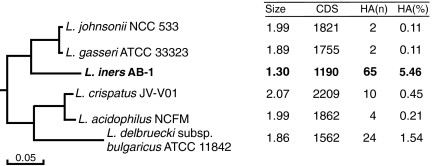
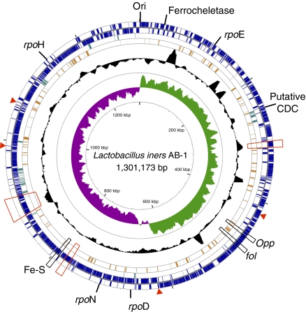
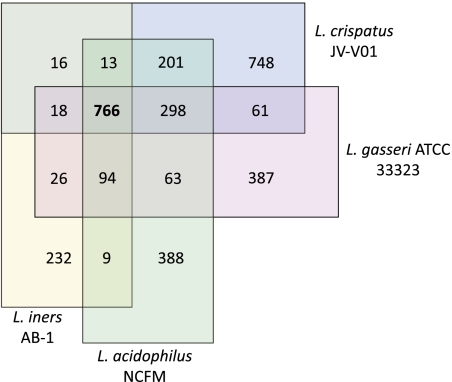
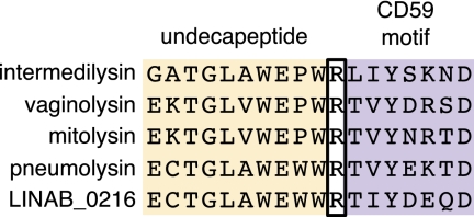
Lactobacilli have long been regarded as important constituents of the healthy human vagina. Lactobacillus iners is the most frequently detected bacterial species in the vagina, but little is known about its characteristics. We report a description of the whole-genome sequence of L. iners AB-1 along with comparative analysis of published genomes of closely related strains of lactobacilli. The genome is the smallest Lactobacillus reported to date, with a 1.3-Mbp single chromosome. The genome seems to have undergone one or more rapid evolution events that resulted in large-scale gene loss and horizontal acquisition of a number of genes for survival in the vagina. L. iners may exhibit specialized adaptation mechanisms to the vaginal environment, such as an iron-sulfur cluster assembly system, and several unique σ factors to regulate gene transcription in this fluctuating environment. A potentially highly expressed homolog of a cholesterol-binding lysin may also contribute to host cell adhesion or act as a defense mechanism against other microbes. Notably, there is a lack of apparent adhesion proteins, but several cell-anchor proteins were identified and may be important for interaction with the host mucosal tissues. L. iners is widely present in healthy females as well as those suffering from bacterial vaginosis or who have undergone antimicrobial therapy, suggesting that it is an important indigenous species of the vagina.


Conflict of interest statement

The authors declare no conflict of interest.

Figures

Fig. 1.
Fig. 1.
Subset of a phylogenetic tree constructed from concatenated ribosomal subunit protein sequences representing the L. iners clade, also part of the acidophilus complex (13) (full figure in SI Appendix, Fig. S1). Key genome features are summarized for each species. Sizes are in millions of base pairs (Mbp). CDS, coding sequences; HA(n), number horizontally acquired genes; HA(%), percent of CDS horizontally acquired.
Fig. 2.
Fig. 2.
Genomic atlas of L. iners AB-1. From the outer circle inward, coding regions are marked on the first two rings: outside the dividing line if encoded on the positive strand and inside the dividing line if encoded on the negative strand. The third ring (dark green) marks ORFs predicted to be horizontally acquired. The fourth ring (orange) shows ORFs predicted to be among the top 10% most highly expressed based on CAI. The fifth ring shows local CG content measured in a sliding window as a black plot. The innermost graph shows the CG skew, with sharp changes in skew occurring at the origin and terminus of replication. Genes of interest, as described in the text, are marked on the outside of the atlas. Regions containing adhesins are marked with red boxes, and single adhesin genes are maked with a red arrow. The atlas was constructed using the CGView Server (70), and a large-scale version can be found in SI Appendix, Fig. S2.
Fig. 3.
Fig. 3.
Venn diagram representing orthologous proteins between select species of the L. iners clade. Values are the number of orthologous genes between overlapping species as predicted by InParanoid (24). The 766 genes conserved between the four species represent a relatively large proportion of the total gene content of L. iners (64%).
Fig. 4.
Fig. 4.
Comparison of the distribution of genes by COG functional category. The white bar represents the mean and SD of the number of genes in each COG functional category for four species of the L. iners clade (L. johnsonii, L. gasseri, L. acidophilus, and L. bulgaricus). The values for L. iners are plotted in the black bar. Marked with asterisks are values in L. iners that are at least 2 SDs away from the mean of the other organisms in the same COG category. COG functional categories: J, translation, ribosomal structure, and biogenesis; K, transcription; L, DNA replication, recombination, and repair; D, cell division and chromosome partitioning; O, posttranslational modification, protein turnover, and chaperones; M, cell envelope biogenesis, outer membrane; N, cell motility and secretion; P, inorganic ion transport and metabolism; T, signal transduction mechanisms; C, energy production and conversion; G, carbohydrate transport and metabolism; E, amino acid transport and metabolism; F, nucleotide transport and metabolism; H, coenzyme metabolism; I, lipid metabolism; Q, secondary metabolites biosynthesis, transport, and catabolism; R, general function prediction only; S, function unknown; V, defense mechanisms; U, intracellular trafficking and secretion.
Fig. 5.
Fig. 5.
Alignment of conserved motifs in the putative cholesterol-dependent cytolysin (LINAB1_0216) with related cytolysins. The cholesterol-binding undecapeptide extends for the first 11 residues. The residues beginning at the R in intermedilysin compose the CD59 motif. Intermedilysin has a fourfold higher affinity to CD59 compared to mitolysin due to the SKN residues present in the CD59 motif compared to the NRT residues present in mitolysin (28). LINAB1_0216 has one or more differences from the intermedilysin in each of the two motifs, the most drastic being the SKN to DEQ difference that changes a polar–basic–polar trio to an acidic–acidic–polar trio.
Fig. 6.
Fig. 6.
Transmission EM of immunogold-labeled L. iners AB-1 (black arrows) in association with human vaginal epithelial cells. Vaginal swab samples collected from healthy women were prepared for EM by thin sectioning. The L. iners cells were labeled with a polyclonal antiserum raised in rabbits against formalin-fixed whole cells of L. iners followed by a secondary goat anti-rabbit gold-conjugated antibody (10-nm gold particles). An even surface distribution of gold particles is expected for L. iners (marked by arrow) but not other bacteria because antibodies not specific to L. iners AB-1 were absorbed out of the serum as described in SI Appendix, SI Text. (Scale bar: 500 nm.)

Similar articles

Cited by

References

    1. Burton JP, Cadieux PA, Reid G. Improved understanding of the bacterial vaginal microbiota of women before and after probiotic instillation. Appl Environ Microbiol. 2003;69:97–101. - PMC - PubMed
    1. Fredricks DN, Fiedler TL, Marrazzo JM. Molecular identification of bacteria associated with bacterial vaginosis. N Engl J Med. 2005;353:1899–1911. - PubMed
    1. Zhou X, et al. The vaginal bacterial communities of Japanese women resemble those of women in other racial groups. FEMS Immunol Med Microbiol. 2010;58:169–181. - PMC - PubMed
    1. Ravel J, et al. Vaginal microbiome of reproductive-age women. Proc Natl Acad Sci USA. 2010:4680–4687. - PMC - PubMed
    1. Hummelen R, et al. Deep sequencing of the vaginal microbiota of women with HIV. PLoS ONE. 2010;5:e12078. - PMC - PubMed

Publication types

Substances