. 2011 Jan;55(1):302-10.
doi: 10.1128/AAC.00633-10. Epub 2010 Nov 8.

Changes in cell wall synthesis and ultrastructure during paradoxical growth effect of caspofungin on four different Candida species

Affiliations

Changes in cell wall synthesis and ultrastructure during paradoxical growth effect of caspofungin on four different Candida species

Fernando C Bizerra et al. Antimicrob Agents Chemother. 2011 Jan.

Abstract

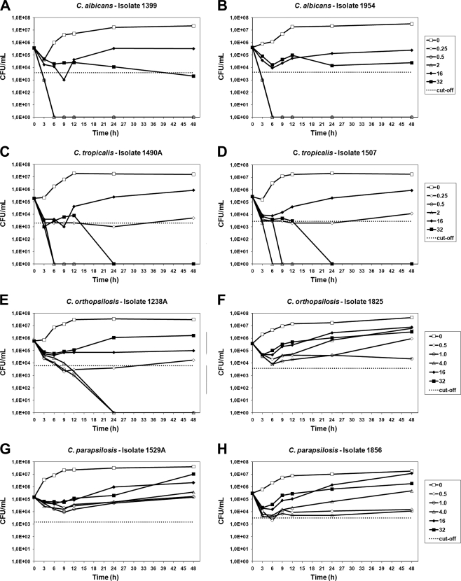
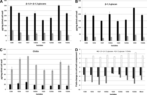
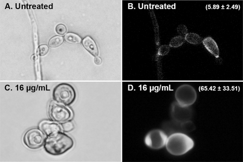
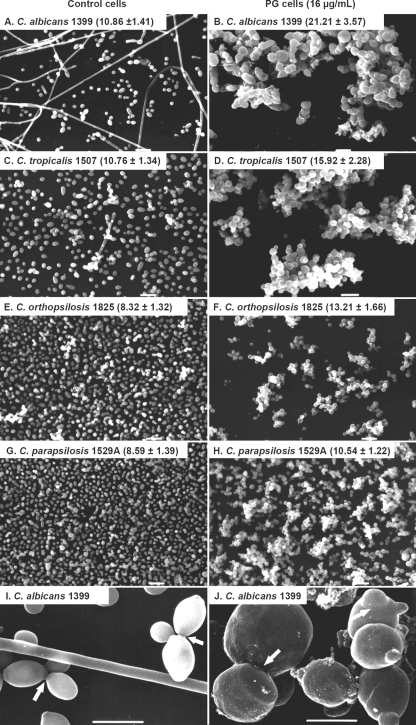
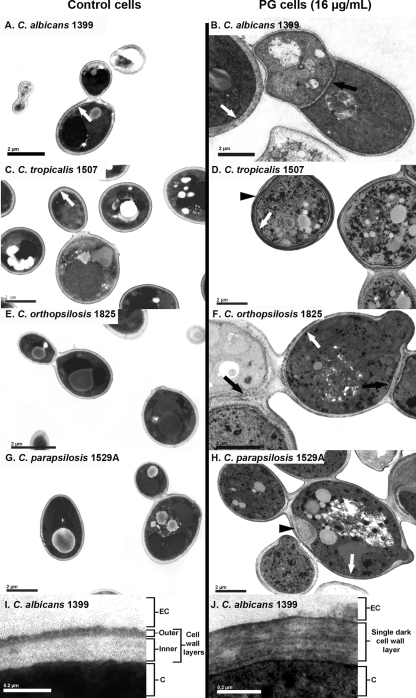
Paradoxical growth (PG) has been described for echinocandins and is characterized by cell growth at drug concentrations above the MIC. In this study, two isolates each of Candida albicans, C. tropicalis, C. orthopsilosis, and C. parapsilosis, all of which displaying PG in response to caspofungin, were subjected to MIC, minimal fungicidal concentration (MFC), and time-kill curve assays to evaluate the levels of PG. Cell wall components and ultrastructural modifications of the PG cells were also investigated. The results showed that when cell growth and survival were evaluated by MFC or time-kill curve assays, high concentrations of caspofungin did not show fungicidal activity against PG cells. Furthermore, for C. parapsilosis and C. orthopsilosis, time-kill curves were more discriminatory than MFCs in detecting the PG effect. The four different Candida species studied demonstrated similar alterations in cell wall components and ultrastructure associated with PG. In PG cells, β-1,3-glucan content decreased from 2.7- to 7.8-fold, whereas chitin content increased from 4.0- to 6.6-fold. An electron microscopy study of the PG cells revealed morphological alterations, clumping of cells, enlarged cells, the absence of filamentation, abnormal septa, and accumulation of chitin in the cell wall. Also, PG cells basically exhibited a single dark high-density layer in the cell wall, indicating the loss of the β-1,3-glucan layer. Our results present novel details about the ultrastructural alterations that occur in C. albicans, C. parapsilosis, C. orthopsilosis, and C. tropicalis during PG and show that chitin is the major component of the cell walls of PG cells. Stimulation of chitin synthesis may represent a rescue mechanism against caspofungin activity.


Figures

FIG. 1.
FIG. 1.
Time-kill curves of all tested Candida isolates: C. albicans (A and B), C. tropicalis (C and D), C. orthopsilosis (E and F), and C. parapsilosis (G and H). CAS concentrations used for each species are shown in the respective graphs. The dotted lines represent a >99% growth reduction compared with the initial inoculum sizes (fungicidal activities).
FIG. 2.
FIG. 2.
Cell wall components of control and PG cells. (A to C) The absolute values of β-1,6- plus β-1,3-glucan (A), β-1,3-glucan (B), and chitin (C) of the control and PG cells (black and gray bars, respectively) were expressed as the number of micrograms of carbohydrate per milligram of dried cell wall. (D) The graph represents the fold change in β-1,6- plus β-1,3-glucan (black), β-1,3-glucan (gray), and chitin (white) contents of PG cells compared to those of control cells.
FIG. 3.
FIG. 3.
Fluorescence microscopy of C. albicans 1399 control (A and B) and PG (C and D) cells stained with calcofluor white (CFW). The value (mean ± standard deviation) of the CFW fluorescence intensity is related to the chitin content of the cell wall (B and D). PG cells showed regions exhibiting the accumulation of chitin and higher fluorescence intensity values than those of control cells (P < 0.05).
FIG. 4.
FIG. 4.
Scanning electron micrographs of Candida strains, comparing control cells and PG cells grown with 16 μg/ml of CAS. The values represent the perimeters of cells (in μm; mean ± standard deviation). PG cells were larger in size than control cells (P < 0.01). Panels I and J show higher magnifications of panels A and B, respectively; the ring around the constriction between mother cell and bud was noted only in control cells (arrows). Scale bars, 10 μm (A to H) and 5 μm (I and J).
FIG. 5.
FIG. 5.
Transmission electron micrographs of Candida strains, comparing control cells and PG cells grown with 16 μg/ml of CAS. Black arrows indicate the abnormal septa in PG cells. White arrows show the cell walls of control cells, which exhibited two layers, with a predominance of the more-electron-dense layer in PG cells. Black arrowheads show a multilayered cell wall (D) and a focal enlargement of the cell wall (H) in PG cells. Panels I and J show higher-magnification electron micrographs of control and PG cells, respectively. (I) As described above, control cells exhibited cell walls with two layers, an electron-dense outer layer and an inner layer of low electron density. (J) In PG cells, a decrease of inner cell wall layers and a predominance of the more-electron-dense layers were observed. EC, extracellular environment; C, cytosol. Scale bars, 2 μm (A to H) and 0.2 μm (I and J).

Similar articles

Cited by

References

    1. Balashov, S. V., S. Park, and D. S. Perlin. 2006. Assessing resistance to the echinocandin antifungal drug caspofungin in Candida albicans by profiling mutations in FKS1. Antimicrob. Agents Chemother. 50:2058-2063. - PMC - PubMed
    1. Barchiesi, F., E. Spreghini, S. Tomassetti, D. Arzeni, D. Giannini, and G. Scalise. 2005. Comparison of the fungicidal activities of caspofungin and amphotericin B against Candida glabrata. Antimicrob. Agents Chemother. 49:4989-4992. - PMC - PubMed
    1. Barchiesi, F., E. Spreghini, S. Tomassetti, A. Della Vittoria, D. Arzeni, E. Manso, and G. Scalise. 2006. Effects of caspofungin against Candida guilliermondii and Candida parapsilosis. Antimicrob. Agents Chemother. 50:2719-2727. - PMC - PubMed
    1. Bizerra, F. C., C. V. Nakamura, C. de Poersch, T. I. Estivalet Svidzinski, R. M. Borsato Quesada, S. Goldenberg, M. A. Krieger, and S. F. Yamada-Ogatta. 2008. Characteristics of biofilm formation by Candida tropicalis and antifungal resistance. FEMS Yeast Res. 8:442-450. - PubMed
    1. Canton, E., J. Peman, M. Romero, A. Valentin, and M. Gobernado. 2007. The fungicidal activity and paradoxical effect of caspofungin against yeast. Influence of culture medium and incubation time. Rev. Esp. Quimioter. 20:433-441. (In Spanish.) - PubMed

Publication types

LinkOut - more resources