. 2011 Jun;21(6):1395-407.
doi: 10.1093/cercor/bhq219. Epub 2010 Nov 8.

The corticofugal neuron-associated genes ROBO1, SRGAP1, and CTIP2 exhibit an anterior to posterior gradient of expression in early fetal human neocortex development

Affiliations

The corticofugal neuron-associated genes ROBO1, SRGAP1, and CTIP2 exhibit an anterior to posterior gradient of expression in early fetal human neocortex development

Bui Kar Ip et al. Cereb Cortex. 2011 Jun.

Abstract

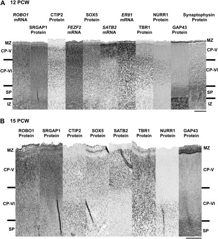
Developing neocortical progenitors express transcription factors in gradients that induce programs of region-specific gene expression. Our previous work identified anteriorly upregulated expression gradients of a number of corticofugal neuron-associated gene probe sets along the anterior-posterior axis of the human neocortex (8-12 postconceptional weeks [PCW]). Here, we demonstrate by real-time polymerase chain reaction, in situ hybridization and immunohistochemistry that 3 such genes, ROBO1, SRGAP1, and CTIP2 are highly expressed anteriorly between 8-12 PCW, in comparison with other genes (FEZF2, SOX5) expressed by Layer V, VI, and subplate neurons. All 3 were prominently expressed by early postmitotic neurons in the subventricular zone, intermediate zone, and cortical plate (CP) from 8 to 10 PCW. Between 12 and 15 PCW expression patterns for ER81 and SATB2 (Layer V), TBR1 (Layer V/VI) and NURR1 (Layer VI) revealed Layer V forming. By 15 PCW, ROBO1 and SRGAP1 expression was confined to Layer V, whereas CTIP2 was expressed throughout the CP anteriorly. We observed ROBO1 and SRGAP1 immunoreactivity in medullary corticospinal axons from 11 PCW onward. Thus, we propose that the coexpression of these 3 markers in the anterior neocortex may mark the early location of the human motor cortex, including its corticospinal projection neurons, allowing further study of their early differentiation.


Figures

Figure 1.
Figure 1.
rtPCR confirmation of gradients of ROBO1, SRGAP1, and CTIP2 expression during 8–12 PCW. Table showingfold changes (A) and a graphical representation (B) of relative expression (NRQ) of ROBO1, SRGAP1, CTIP2, FEZF2, and SOX5 determined by rtPCR from RNA extracted from anterior and posterior regions of developing human neocortex aged between 8 and 12 PCW (n = 11). ROBO1 and CTIP2 exhibited statistically significant, large fold changes, indicating a high anterior to low posterior gradient. SRGAP1 showed a small but significant fold change corresponding to a high anterior to low posterior gradient. FEZF2 and SOX5 exhibited high posterior, low anterior gradients of small but significant fold changes. *P < 0.05 (paired Student’s T-test); A, anterior; P, posterior.
Figure 2.
Figure 2.
ROBO1, SRGAP1, and CTIP2 gradients in sections from the developing human neocortex. ISH of sagittal sections for ROBO1, CTIP2, FEZF2 and SOX5 and IHC for SRGAP1 (A, B) revealed that ROBO1, SRGAP1, and CTIP2 were expressed at high levels anteriorly within the SVZ, IZ, and CP between 8–10 PCW. A high posterior, low anterior expression of FEZF2 was detected but at 9 and 10 PCW only. No gradient was detected for SOX5 at any stage investigated. Adjacent sections were selected for each gene/protein at each developmental stage. Higher magnification images, outlined with boxes, were taken at the anterior (At) and posterior regions (Pt) of the neocortex. (C) The optical density of histological staining for ROBO1, CTIP2, FEZF2, SOX5 mRNA and SRGAP1-immunoreactivity at the anterior-most neocortex (gray bars) was measured in the CP and expressed relative to background staining in the VZ (see Materials and Methods) and compared with the same ratios measured at the posterior pole (black bars). *P < 0.05 (paired Student’s T-test). Scale bars represent 200 μm.
Figure 3.
Figure 3.
Laminar localization of ROBO1, SRGAP1, and CTIP2 during early human neocortical development (8–10 PCW). ISH and IHC on paraffin sections revealed expression of ROBO1 mRNA and protein, SRGAP1 protein and CTIP2 mRNA and protein in the SVZ, IZ/SP, and CP between 8 and 10 PCW (A, B). All sections were taken from the dorsolateral region of the neocortex, and adjacent sections were selected for each gene/protein at each developmental stage. Note the higher expression of ROBO1 and SRGAP1 protein in the IZ and SP compared with ROBO1 mRNA, which was more highly expressed in the CP. This suggests a predominant localization of the proteins to receptor complexes in axons compared with the predominant localization of mRNA in the cell body. CTIP2, a transcription factor, was predominantly localized to the nucleus of positive cells. Scale bars represent 200 μm.
Figure 4.
Figure 4.
Laminar localization of ROBO1, SRGAP1, and CTIP2 at 12 and 15 PCW. (A) Comparison of expression of ROBO1, SRGAP1, and CTIP2 with various laminar-specific markers (SATB2, ER81, TBR1, NURR1 and GAP43 and Synaptophysin) revealed the emergence of Layer V by 12 PCW. The CP was identified by GAP43- and Synaptophysin-negative immunoreactivity, proximal to the MZ. Within the CP, strong SATB2 and ER81 expression was detected in the Layer V above the TBR1- and NURR1-expressing Layer VI. ROBO1, SRGAP1, and CTIP2 together with FEZF2 and SOX5 expression were observed throughout the CP (Layer V and VI). (B) At 15 PCW, NURR1-expressing cells were observed in Layer VI and also in the upper parts of the GAP43-positive SP. Expression of CTIP2 together with SOX5, SATB2, and TBR1 were detected in both Layer V and VI with different intensities, whereas intense immunostaining of ROBO1 and SRGAP1 were observed mostly in the Layer V. All sections were taken from the dorsolateral region of the neocortex, and adjacent sections were selected for each gene/protein at each foetal stage. Scale bars represent 200 μm.
Figure 5.
Figure 5.
Cellular localization of ROBO1, SRGAP1, and CTIP2 from 10 to 12 PCW. (A) At 10 PCW, within the SVZ, most CTIP2-positive cells (green) did not express TBR2 (red) a marker for INPs, indicating that CTIP2 positive cells are predominantly postmitotic neurons. (B) At 12 PCW, within Layer V of the CP, postmitotic neurons predominantly expressed either CTIP2 (green) or SATB2 (red), and very few coexpressed both markers (arrow). However, there were many more ROBO1 and SATB2 coexpressing cells (arrow). (CE) At 10 PCW, within the CP, a subset of postmitotic neurons coexpressed CTIP2, ROBO1, and SRGAP1 (arrows). Scale bars represent 100 μm.
Figure 6.
Figure 6.
ROBO1 and SRGAP1 are CST markers. (A-C) Show caudal medulla sections at the level of olives at 11 PCW. GAP43 expression was detected in all growing axons of different pathways revealed in this section (A). However, ROBO1- (B) and SRGAP1- (C) immunoreactive fibers were observed only in the 2 small regions at the ventral surface of the medulla, presumably where the future medullary pyramids are formed. (DF) Show sections of caudal medulla at the level of the decussation, at 11 PCW. Small numbers of fibers crossing over the midline in the medulla were immunoreactive for GAP43 (D) but negative for ROBO1 (E) and SRGAP1 (F). (GJ) Show caudal medulla sections at the level of decussation at 14 and 17 PCW. Both ROBO1 (G, I) and SRGAP1 (H, J) were expressed in fibers crossing over at the decussation. Scale bars represent 500 μm.

Similar articles

Cited by

References

    1. Abrahams BS, Tentler D, Perederiy JV, Oldham MC, Coppola G, Geschwind DH. Genome-wide analyses of human perisylvian cerebral cortical patterning. Proc Natl Acad Sci U S A. 2007;104:17849–17854. - PMC - PubMed
    1. Alcamo EA, Chirivella L, Dautzenberg M, Dobreva G, Farinas I, Grosschedl R, McConnell SK. Satb2 regulates callosal projection neuron identity in the developing cerebral cortex. Neuron. 2008;57:364–377. - PubMed
    1. Andrews W, Barber M, Hernadez-Miranda LR, Xian J, Rakic S, Sundaresan V, Rabbitts TH, Pannell R, Rabbitts P, Thompson H, et al. The role of Slit-Robo signaling in the generation, migration and morphological differentiation of cortical interneurons. Dev Biol. 2008;313:648–658. - PubMed
    1. Andrews W, Liapi A, Plachez C, Camurri L, Zhang J, Mori S, Murakami F, Parnavelas JG, Sundaresan V, Richards LJ. Robo1 regulates the development of major axon tracts and interneuron migration in the forebrain. Development. 2006;133:2243–2252. - PubMed
    1. Andrews WD, Barber M, Parnavelas JG. Slit-Robo interactions during cortical development. J Anat. 2007;211:188–198. - PMC - PubMed

Publication types

MeSH terms