Cholinergic and non-cholinergic projections from the pedunculopontine and laterodorsal tegmental nuclei to the medial geniculate body in Guinea pigs
- PMID: 21060717
- PMCID: PMC2972721
- DOI: 10.3389/fnana.2010.00137
Cholinergic and non-cholinergic projections from the pedunculopontine and laterodorsal tegmental nuclei to the medial geniculate body in Guinea pigs
Abstract
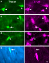
The midbrain tegmentum is the source of cholinergic innervation of the thalamus and has been associated with arousal and control of the sleep/wake cycle. In general, the innervation arises bilaterally from the pedunculopontine tegmental nucleus (PPT) and the laterodorsal tegmental nucleus (LDT). While this pattern has been observed for many thalamic nuclei, a projection from the LDT to the medial geniculate body (MG) has been questioned in some species. We combined retrograde tracing with immunohistochemistry for choline acetyltransferase (ChAT) to identify cholinergic projections from the brainstem to the MG in guinea pigs. Double-labeled cells (retrograde and immunoreactive for ChAT) were found in both the PPT (74%) and the LDT (26%). In both nuclei, double-labeled cells were more numerous on the ipsilateral side. About half of the retrogradely labeled cells were immunonegative, suggesting they are non-cholinergic. The distribution of these immunonegative cells was similar to that of the immunopositive ones: more were in the PPT than the LDT and more were on the ipsilateral than the contralateral side. The results indicate that both the PPT and the LDT project to the MG, and suggest that both cholinergic and non-cholinergic cells contribute substantially to these projections.
Keywords: GABA; acetylcholine; arousal; auditory; glutamate; sleep; thalamus.
Figures


Similar articles
-
Cholinergic cells of the pontomesencephalic tegmentum: connections with auditory structures from cochlear nucleus to cortex.Hear Res. 2011 Sep;279(1-2):85-95. doi: 10.1016/j.heares.2010.12.019. Epub 2010 Dec 30. Hear Res. 2011. PMID: 21195150 Free PMC article. Review.
-
Sources of cholinergic input to the inferior colliculus.Neuroscience. 2009 Apr 21;160(1):103-14. doi: 10.1016/j.neuroscience.2009.02.036. Epub 2009 Mar 10. Neuroscience. 2009. PMID: 19281878 Free PMC article.
-
Projections from auditory cortex to midbrain cholinergic neurons that project to the inferior colliculus.Neuroscience. 2010 Mar 10;166(1):231-40. doi: 10.1016/j.neuroscience.2009.12.008. Epub 2009 Dec 13. Neuroscience. 2010. PMID: 20005923 Free PMC article.
-
Ascending projections from the pedunculopontine tegmental nucleus and the adjacent mesopontine tegmentum in the rat.J Comp Neurol. 1988 Aug 22;274(4):483-515. doi: 10.1002/cne.902740403. J Comp Neurol. 1988. PMID: 2464621
-
Paradoxical sleep and its chemical/structural substrates in the brain.Neuroscience. 1991;40(3):637-56. doi: 10.1016/0306-4522(91)90002-6. Neuroscience. 1991. PMID: 2062436 Review.
Cited by
-
Multiple origins of cholinergic innervation of the cochlear nucleus.Neuroscience. 2011 Apr 28;180:138-47. doi: 10.1016/j.neuroscience.2011.02.010. Epub 2011 Feb 12. Neuroscience. 2011. PMID: 21320579 Free PMC article.
-
Cholinergic cells of the pontomesencephalic tegmentum: connections with auditory structures from cochlear nucleus to cortex.Hear Res. 2011 Sep;279(1-2):85-95. doi: 10.1016/j.heares.2010.12.019. Epub 2010 Dec 30. Hear Res. 2011. PMID: 21195150 Free PMC article. Review.
-
Cholinergic brain network deficits associated with vestibular sensory conflict deficits in Parkinson's disease: correlation with postural and gait deficits.J Neural Transm (Vienna). 2022 Aug;129(8):1001-1009. doi: 10.1007/s00702-022-02523-3. Epub 2022 Jun 26. J Neural Transm (Vienna). 2022. PMID: 35753016 Free PMC article.
-
Subcollicular projections to the auditory thalamus and collateral projections to the inferior colliculus.Front Neuroanat. 2014 Jul 18;8:70. doi: 10.3389/fnana.2014.00070. eCollection 2014. Front Neuroanat. 2014. PMID: 25100950 Free PMC article.
-
Mechanisms of GABAergic and cholinergic neurotransmission in auditory thalamus: Impact of aging.Hear Res. 2021 Mar 15;402:108003. doi: 10.1016/j.heares.2020.108003. Epub 2020 Jun 11. Hear Res. 2021. PMID: 32703637 Free PMC article. Review.
References
Grants and funding
LinkOut - more resources
Full Text Sources

