

What have We Learned from "Perturbing" the Human Cortical Motor System with Transcranial Magnetic Stimulation?
- PMID: 21060721
- PMCID: PMC2972749
- DOI: 10.3389/fnhum.2010.00173
What have We Learned from "Perturbing" the Human Cortical Motor System with Transcranial Magnetic Stimulation?
Abstract
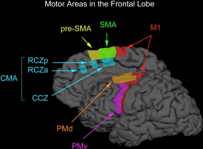
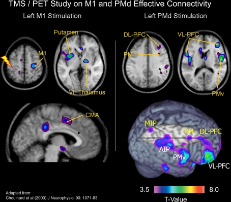
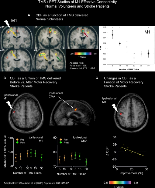
The purpose of this paper is twofold. First, we will review different approaches that one can use with transcranial magnetic stimulation (TMS) to study both its effects on motor behavior and on neural connections in the human brain. Second, we will present evidence obtained in TMS-based studies showing that the dorsal premotor area (PMd), the ventral premotor area (PMv), the supplementary motor area (SMA), and the pre-supplementary motor area (pre-SMA) each have different roles to play in motor behavior. We highlight the importance of the PMd in response selection based on arbitrary cues and in the control of arm movements, the PMv in grasping and in the discrimination of bodily actions, the SMA in movement sequencing and in bimanual coordination, and the pre-SMA in cognitive control. We will also discuss ways in which TMS can be used to chart "true" cerebral reorganization in clinical populations and how TMS might be used as a therapeutic tool to facilitate motor recovery after stroke. We will end our review by discussing some of the methodological challenges and future directions for using this tool in basic and clinical neuroscience.
Keywords: effective connectivity; functional connectivity; functional neuroimaging; motor system; premotor area; stroke recovery; supplementary motor area; transcranial magnetic stimulation.
Figures

Similar articles
-
Assessing the effective connectivity of premotor areas during real vs imagined grasping: a DCM-PEB approach.Neuroimage. 2021 Apr 15;230:117806. doi: 10.1016/j.neuroimage.2021.117806. Epub 2021 Jan 29. Neuroimage. 2021. PMID: 33524574
-
The Topography of Visually Guided Grasping in the Premotor Cortex: A Dense-Transcranial Magnetic Stimulation (TMS) Mapping Study.J Neurosci. 2020 Aug 26;40(35):6790-6800. doi: 10.1523/JNEUROSCI.0560-20.2020. Epub 2020 Jul 24. J Neurosci. 2020. PMID: 32709693 Free PMC article.
-
Influence of the supplementary motor area on primary motor cortex excitability during movements triggered by neutral or emotionally unpleasant visual cues.Exp Brain Res. 2003 Mar;149(2):214-21. doi: 10.1007/s00221-002-1346-8. Epub 2003 Jan 25. Exp Brain Res. 2003. PMID: 12610690
-
Transcranial Magnetic Stimulation in Tourette Syndrome: A Historical Perspective, Its Current Use and the Influence of Comorbidities in Treatment Response.Brain Sci. 2018 Jul 6;8(7):129. doi: 10.3390/brainsci8070129. Brain Sci. 2018. PMID: 29986411 Free PMC article. Review.
-
A Systematic Review of Integrated Functional Near-Infrared Spectroscopy (fNIRS) and Transcranial Magnetic Stimulation (TMS) Studies.Front Neurosci. 2019 Feb 28;13:84. doi: 10.3389/fnins.2019.00084. eCollection 2019. Front Neurosci. 2019. PMID: 30872985 Free PMC article.
Cited by
-
Hierarchical vector auto-regressive models and their applications to multi-subject effective connectivity.Front Comput Neurosci. 2013 Nov 12;7:159. doi: 10.3389/fncom.2013.00159. eCollection 2013. Front Comput Neurosci. 2013. PMID: 24282401 Free PMC article.
-
Resting State Functional Connectivity Signatures of MRgFUS Vim Thalamotomy in Parkinson's Disease: A Preliminary Study.Front Neurol. 2022 Jan 12;12:786734. doi: 10.3389/fneur.2021.786734. eCollection 2021. Front Neurol. 2022. PMID: 35095731 Free PMC article.
-
Reduced Performance During a Sentence Repetition Task by Continuous Theta-Burst Magnetic Stimulation of the Pre-supplementary Motor Area.Front Neurosci. 2018 May 29;12:361. doi: 10.3389/fnins.2018.00361. eCollection 2018. Front Neurosci. 2018. PMID: 29896086 Free PMC article.
-
The role of areas MT+/V5 and SPOC in spatial and temporal control of manual interception: an rTMS study.Front Behav Neurosci. 2013 Mar 5;7:15. doi: 10.3389/fnbeh.2013.00015. eCollection 2013. Front Behav Neurosci. 2013. PMID: 23468002 Free PMC article.
-
The supplementary motor area modulates interhemispheric interactions during movement preparation.Hum Brain Mapp. 2019 May;40(7):2125-2142. doi: 10.1002/hbm.24512. Epub 2019 Jan 17. Hum Brain Mapp. 2019. PMID: 30653778 Free PMC article.
References
-
- Ameli M., Grefkes C., Kemper F., Riegg F. P., Rehme A. K., Karbe H., Fink G. R., Nowak D. A. (2009). Differential effects of high-frequency repetitive transcranial magnetic stimulation over ipsilesional primary motor cortex in cortical and subcortical middle cerebral artery stroke. Ann. Neurol. 66, 298–30910.1002/ana.21725 - DOI - PubMed
LinkOut - more resources
Full Text Sources