. 2010 Nov 9;1(5):e00268-10.
doi: 10.1128/mBio.00268-10.

Head-to-tail intramolecular interaction of herpes simplex virus type 1 regulatory protein ICP27 is important for its interaction with cellular mRNA export receptor TAP/NXF1

Affiliations

Head-to-tail intramolecular interaction of herpes simplex virus type 1 regulatory protein ICP27 is important for its interaction with cellular mRNA export receptor TAP/NXF1

Felicia P Hernandez et al. mBio. .

Abstract

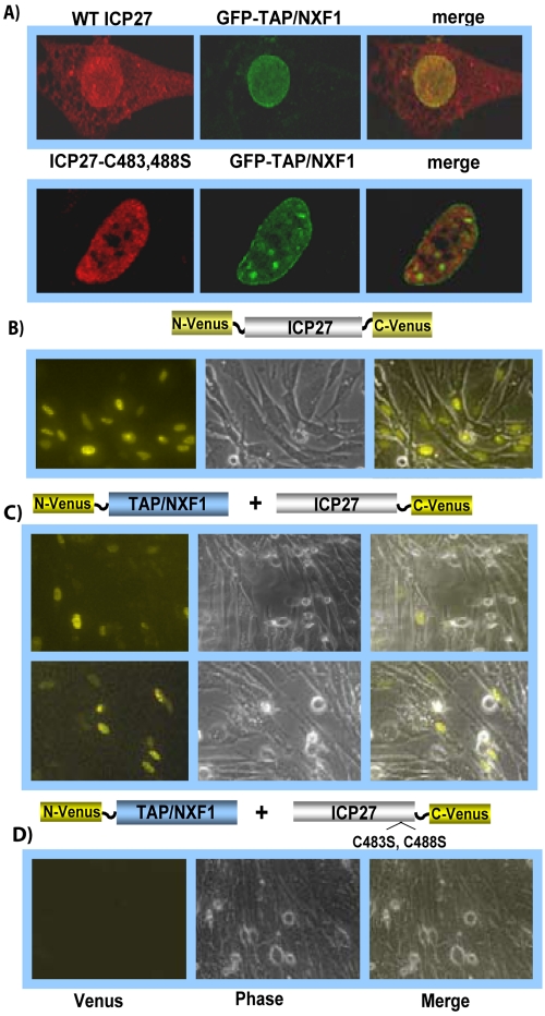
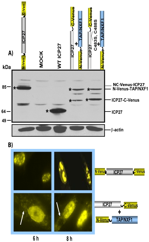
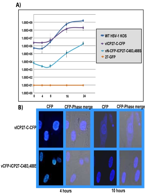
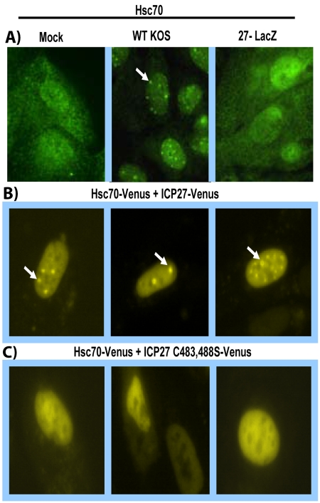
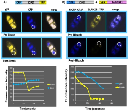
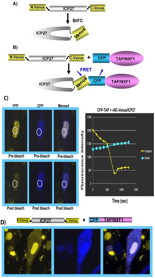
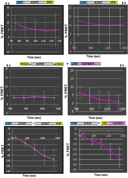
Herpes simplex virus type 1 (HSV-1) protein ICP27 has many important functions during infection that are achieved through interactions with a number of cellular proteins. In its role as a viral RNA export protein, ICP27 interacts with TAP/NXF1, the cellular mRNA export receptor, and both the N and C termini of ICP27 must be intact for this interaction to take place. Here we show by bimolecular fluorescence complementation (BiFC) that ICP27 interacts directly with TAP/NXF1 during infection, and this interaction failed to occur with an ICP27 mutant bearing substitutions of serines for cysteines at positions 483 and 488 in the C-terminal zinc finger. Recently, we showed that ICP27 undergoes a head-to-tail intramolecular interaction, which could make the N- and C-terminal regions accessible for binding to TAP/NXF1. To determine the importance of intramolecular association of ICP27 to its interaction with TAP/NXF1, we performed BiFC-based fluorescence resonance energy transfer (FRET) by acceptor photobleaching. BiFC-based FRET showed that the interaction between ICP27 and TAP/NXF1 occurred in living cells upon head-to-tail intramolecular association of ICP27, further establishing that TAP/NXF1 interacts with both the N and C termini of ICP27.


Figures

FIG 1
FIG 1
ICP27 interacts directly with TAP/NXF1. (A) RSF were cotransfected with GFP-TAP/NXF1 DNA and with ICP27 constructs carrying wild-type ICP27 or ICP27-C483,488S and were subsequently infected with 27-LacZ for 8 h. Immunofluorescent staining was performed with anti-ICP27 antibody, and GFP fluorescence was viewed directly. (B to D) Cells were transfected with NC-Venus-ICP27 (B) or were cotransfected with N-Venus–TAP/NXF1 and ICP27–C-Venus (C) or with N-Venus–TAP/NXF1 and ICP27-C483,488S–C-Venus (D). Twenty-four hours later, cells were infected with 27-LacZ for 8 h. Cells were viewed directly for Venus fluorescence using a Zeiss LSM 510 Meta confocal microscope at a magnification of ×20.
FIG 2
FIG 2
Western blot analysis of TAP/NXF1-Venus and ICP27-Venus fusion proteins. (A) RSF were transfected with plasmid DNA encoding the proteins indicated and infected with 27-LacZ to induce expression of ICP27 constructs. Cells were harvested 8 h after infection, and proteins were fractionated on a 10% SDS-polyacrylamide gel and transferred to nitrocellulose. Membranes were probed with antibodies against GFP, ICP27, and β-actin as described previously (27). Asterisks mark the protein bands. (B) RSF were transfected with NC-Venus-ICP27 or cotransfected with ICP27–C-Venus and N-Venus–TAP/NXF1 and later infected with 27-LacZ for 6 and 8 h, as indicated. Venus fluorescence was viewed directly using a Zeiss LSM 510 Meta confocal microscope at a magnification of ×63. White arrows point to Venus cytoplasmic fluorescence.
FIG 3
FIG 3
CFP–ICP27-C483,488S is confined to the nucleus during infection. (A) Vero cells were infected with HSV-1 KOS, 27-GFP, vICP27-C–CFP, and vN-CFP–ICP27-C483,488S at an MOI of 1. Experiments were performed in triplicate, and virus was harvested at 0, 4, 8, 16, and 24 h after infection. Plaque assays were performed in duplicate on Vero cells. (B) RSF were infected with vICP27-C–CFP or vN-CFP–ICP27-C483,488S at an MOI of 10 for 4 and 10 h after infection. CFP fluorescence was viewed with a Zeiss LSM 510 Meta confocal microscope at a magnification of ×63.
FIG 4
FIG 4
Hsc70 interacts directly with ICP27. RSF were cotransfected with N-Venus–Hsc70 and ICP27–C-Venus or with N-Venus–Hsc70 and ICP27-C483,488S–C-Venus as depicted and were subsequently infected with 27-LacZ for 6 h (A) or 8 h (B). Venus fluorescence was viewed with a Zeiss LSM 510 Meta confocal microscope at a magnification of ×20.
FIG 5
FIG 5
Mutation of the ICP27 zinc finger affects the functional interaction of ICP27 and Hsc70. (A) RSF were either mock infected or infected with HSV-1 KOS or 27-LacZ as indicated for 6 h. Cells were fixed and stained with anti-Hsc70 antibody as described (5). (B, C) RSF were cotransfected with N-Venus–Hsc70 and ICP27–C-Venus (B) or with ICP27-C483,488S–C-Venus (C) as indicated and infected with 27-LacZ. At 8 h after infection, Venus fluorescence was viewed using a Zeiss LSM 510 Meta confocal microscope at a magnification of ×63. White arrows point to Hsc70 nuclear foci.
FIG 6
FIG 6
Interaction between CFP-ICP27 and YFP-TAP/NXF1 was not observed by FRET after acceptor photobleaching. (A) RSF were transfected with CFP-ICP27-YFP and were infected with 27-LacZ for 6 h. Merged images depicting the colocalization of YFP-ICP27 and CFP-ICP27 are shown, with the regions of interest for donor and acceptor images before and after bleaching circled. Quantification of FRET for the circled regions is displayed graphically as fluorescence intensity over time. (B) RSF were transfected with CFP-ICP27 and YFP-ICP27 and were then infected with 27-LacZ for 6 h. Merged images depicting the colocalization of CFP-ICP27 and YFP-TAP/NXF1 are shown, with the regions of interest for bleaching circled. Quantification of FRET for the circled regions is displayed graphically as fluorescence intensity over time, as described in Materials and Methods. FRET analysis was performed by using an LSM confocal microscope at a magnification of ×63.
FIG 7
FIG 7
BiFC-FRET after photobleaching demonstrates the interaction of TAP/NXF1 with ICP27 upon head-to-tail association. (A) Schematic representation of BiFC for NC-Venus-ICP27. (B) Model showing FRET between CFP-TAP/NXF1 and NC-Venus-ICP27. (C) RSF were transfected with CFP-TAP/NXF1 and were subsequently infected with vNC-Venus-ICP27 at an MOI of 10. Merged images depicting the colocalization of ICP27 with TAP/NXF1 are shown, with the regions of interest circled before and after bleaching of the acceptor. Quantification of FRET for the circled regions is displayed graphically as fluorescence intensity over time. FRET analysis was performed as described in the legend to Fig. 6. (D) RSF were transfected with CFP-TAP/NXF1 and were subsequently infected with vNC-Venus-ICP27 for 6 h. Venus and CFP fluorescence were viewed directly using an LSM confocal microscope at a magnification of ×63.
FIG 8
FIG 8
Analysis of percent FRET over time. RSF were transfected with the constructs indicated and were subsequently infected with 27-LacZ or with vNC-Venus-ICP27, as indicated. FRET after acceptor photobleaching was performed by photobleaching the acceptor protein Venus or YFP at a specific location within the cell using the 514-nm laser line at 100% transmission. The change in donor fluorescence was quantified by comparing prebleach and postbleach levels of CFP fluorescence from the images. The FRET efficiency (EF) obtained from at least 30 different bleached regions of interest was calculated as described in Materials and Methods. FRET efficiency is shown as percent FRET over time. Experiments were performed in triplicate. Error bars are shown.

Similar articles

Cited by

References

    1. Bryant H. E., Wadd S., Lamond A. I., Silverstein S. J., Clements J. B. 2001. Herpes simplex virus IE63 (ICP27) protein interacts with spliceosome-associated protein 145 and inhibits splicing prior to the first catalytic step. J. Virol. 75:4376–4385 - PMC - PubMed
    1. Sciabica K. S., Dai Q. J., Sandri-Goldin R. M. 2003. ICP27 interacts with SRPK1 to mediate HSV-1 inhibition of pre-mRNA splicing by altering SR protein phosphorylation. EMBO J. 22:1608–1619 - PMC - PubMed
    1. Chen I. B., Li L., Silva L., Sandri-Goldin R. M. 2005. ICP27 recruits Aly/REF but not TAP/NXF1 to herpes simplex virus type 1 transcription sites although TAP/NXF1 is required for ICP27 export. J. Virol. 79:3949–3961 - PMC - PubMed
    1. Chen I. B., Sciabica K. S., Sandri-Goldin R. M. 2002. ICP27 interacts with the export factor Aly/REF to direct herpes simplex virus 1 intronless RNAs to the TAP export pathway. J. Virol. 76:12877–12889 - PMC - PubMed
    1. Li L., Johnson L. A., Dai-Ju J. Q., Sandri-Goldin R. M. 2008. Hsc70 focus formation at the periphery of HSV-1 transcription sites requires ICP27. PLoS One 3:e1491 - PMC - PubMed

Publication types

MeSH terms

Substances