

Mapping of genetic abnormalities of primary tumours from metastatic CRC by high-resolution SNP arrays
- PMID: 21060790
- PMCID: PMC2966422
- DOI: 10.1371/journal.pone.0013752
Mapping of genetic abnormalities of primary tumours from metastatic CRC by high-resolution SNP arrays
Abstract
Background: For years, the genetics of metastatic colorectal cancer (CRC) have been studied using a variety of techniques. However, most of the approaches employed so far have a relatively limited resolution which hampers detailed characterization of the common recurrent chromosomal breakpoints as well as the identification of small regions carrying genetic changes and the genes involved in them.
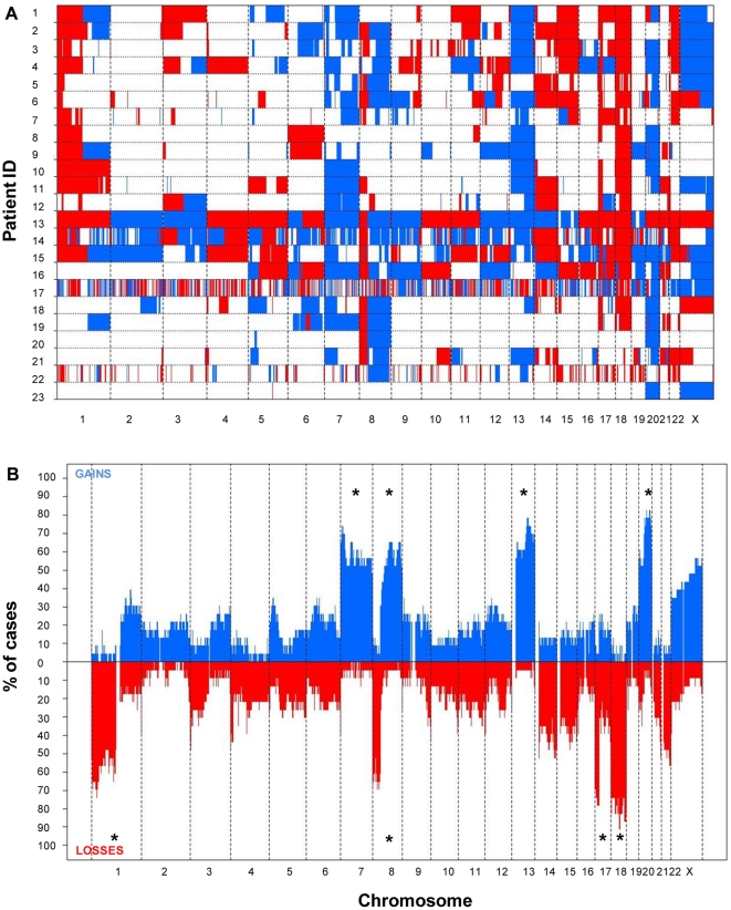
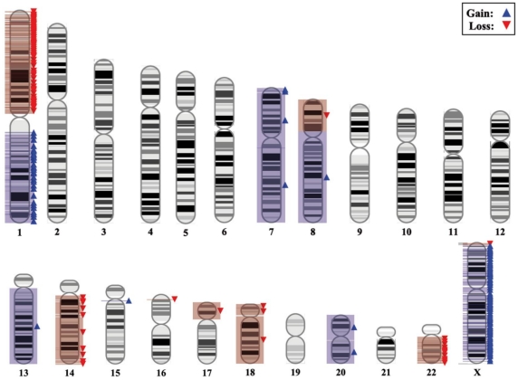
Methodology/principal findings: Here we applied 500K SNP arrays to map the most common chromosomal lesions present at diagnosis in a series of 23 primary tumours from sporadic CRC patients who had developed liver metastasis. Overall our results confirm that the genetic profile of metastatic CRC is defined by imbalanced gains of chromosomes 7, 8q, 11q, 13q, 20q and X together with losses of the 1p, 8p, 17p and 18q chromosome regions. In addition, SNP-array studies allowed the identification of small (<1.3 Mb) and extensive/large (>1.5 Mb) altered DNA sequences, many of which contain cancer genes known to be involved in CRC and the metastatic process. Detailed characterization of the breakpoint regions for the altered chromosomes showed four recurrent breakpoints at chromosomes 1p12, 8p12, 17p11.2 and 20p12.1; interestingly, the most frequently observed recurrent chromosomal breakpoint was localized at 17p11.2 and systematically targeted the FAM27L gene, whose role in CRC deserves further investigations.
Conclusions/significance: In summary, in the present study we provide a detailed map of the genetic abnormalities of primary tumours from metastatic CRC patients, which confirm and extend on previous observations as regards the identification of genes potentially involved in development of CRC and the metastatic process.
Conflict of interest statement
Figures
Similar articles
-
Unique genetic profile of sporadic colorectal cancer liver metastasis versus primary tumors as defined by high-density single-nucleotide polymorphism arrays.Mod Pathol. 2012 Apr;25(4):590-601. doi: 10.1038/modpathol.2011.195. Epub 2012 Jan 6. Mod Pathol. 2012. PMID: 22222638
-
Intratumoural cytogenetic heterogeneity of sporadic colorectal carcinomas suggests several pathways to liver metastasis.J Pathol. 2010 Jul;221(3):308-19. doi: 10.1002/path.2712. J Pathol. 2010. PMID: 20527024
-
-8p12-23 and +20q are predictors of subtypes and metastatic pathways in colorectal cancer: construction of tree models using comparative genomic hybridization data.OMICS. 2011 Jan-Feb;15(1-2):37-47. doi: 10.1089/omi.2010.0101. Epub 2010 Dec 31. OMICS. 2011. PMID: 21194300
-
Chromosomal aberrations related to metastasis of human solid tumors.World J Gastroenterol. 2002 Oct;8(5):769-76. doi: 10.3748/wjg.v8.i5.769. World J Gastroenterol. 2002. PMID: 12378613 Free PMC article. Review.
-
Molecular cytogenetic characterization of pancreas cancer cell lines reveals high complexity chromosomal alterations.Cytogenet Genome Res. 2007;118(2-4):148-56. doi: 10.1159/000108295. Cytogenet Genome Res. 2007. PMID: 18000365 Review.
Cited by
-
Intra-patient Inter-metastatic Genetic Heterogeneity in Colorectal Cancer as a Key Determinant of Survival after Curative Liver Resection.PLoS Genet. 2016 Jul 29;12(7):e1006225. doi: 10.1371/journal.pgen.1006225. eCollection 2016 Jul. PLoS Genet. 2016. PMID: 27472274 Free PMC article.
-
Metastasis suppressor genes.Histol Histopathol. 2013 Mar;28(3):285-92. doi: 10.14670/HH-28.285. Histol Histopathol. 2013. PMID: 23348381 Free PMC article. Review.
-
Endoluminal tumor implant of a colorectal cancer in an anal fistula detected by FISH techniques: a case report.J Gastrointest Oncol. 2021 Apr;12(2):900-905. doi: 10.21037/jgo-20-281. J Gastrointest Oncol. 2021. PMID: 34012678 Free PMC article.
-
Prognostic Impact of EGFR Amplification and Visceral Pleural Invasion in Early Stage Pulmonary Squamous Cell Carcinomas Patients after Surgical Resection of Primary Tumor.Cancers (Basel). 2022 Apr 27;14(9):2174. doi: 10.3390/cancers14092174. Cancers (Basel). 2022. PMID: 35565304 Free PMC article.
-
A comprehensive characterization of genome-wide copy number aberrations in colorectal cancer reveals novel oncogenes and patterns of alterations.PLoS One. 2012;7(7):e42001. doi: 10.1371/journal.pone.0042001. Epub 2012 Jul 31. PLoS One. 2012. PMID: 22860045 Free PMC article.
References
-
- Tsai MS, Su YH, Ho MC, Liang JT, Chen TP, et al. Clinicopathological features and prognosis in resectable synchronous and metachronous colorectal liver metastasis. Ann Surg Oncol. 2007;14:786–94. - PubMed
-
- Rigola MA, Casadevall C, Bernues M, Caballin MR, Fuster C, et al. Analysis of kidney tumors by comparative genomic hybridization and conventional cytogenetics. Cancer Genet Cytogenet. 2002;137:49–53. - PubMed
-
- Garcia J, Duran A, Tabernero MD, Garcia PA, Flores CT, et al. Numerical abnormalities of chromosomes 17 and 18 in sporadic colorectal cancer: Incidence and correlation with clinical and biological findings and the prognosis of the disease. Cytometry B Clin Cytom. 2003;51:14–20. - PubMed
