. 2010 Oct 28;6(10):e1001162.
doi: 10.1371/journal.ppat.1001162.

Nuclear export and import of human hepatitis B virus capsid protein and particles

Affiliations

Nuclear export and import of human hepatitis B virus capsid protein and particles

Hung-Cheng Li et al. PLoS Pathog. .

Abstract

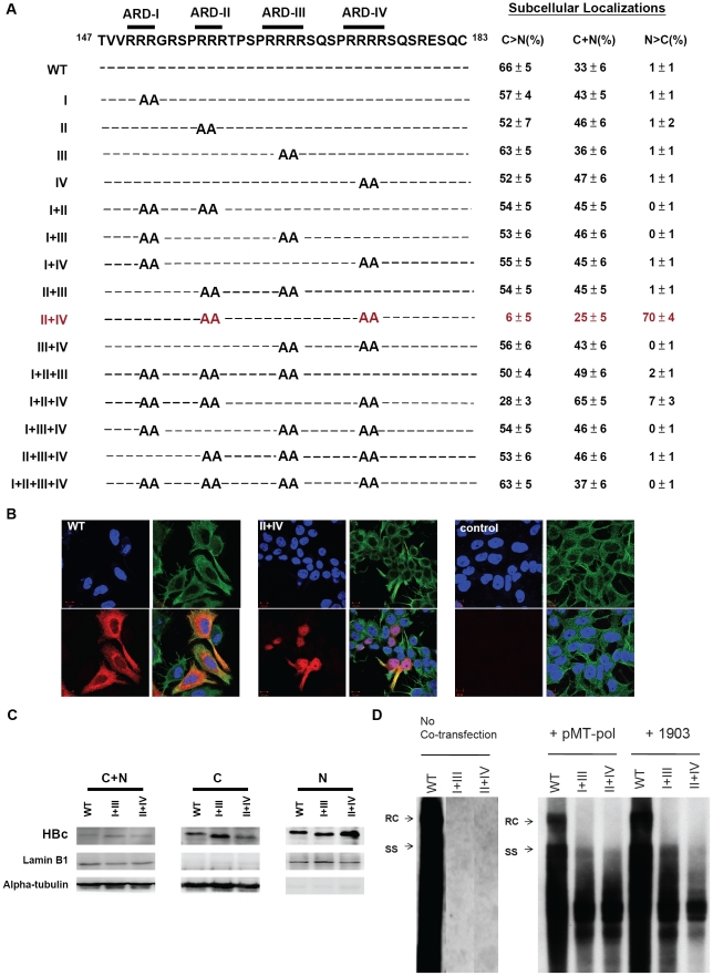
It remains unclear what determines the subcellular localization of hepatitis B virus (HBV) core protein (HBc) and particles. To address this fundamental issue, we have identified four distinct HBc localization signals in the arginine rich domain (ARD) of HBc, using immunofluorescence confocal microscopy and fractionation/Western blot analysis. ARD consists of four tight clustering arginine-rich subdomains. ARD-I and ARD-III are associated with two co-dependent nuclear localization signals (NLS), while ARD-II and ARD-IV behave like two independent nuclear export signals (NES). This conclusion is based on five independent lines of experimental evidence: i) Using an HBV replication system in hepatoma cells, we demonstrated in a double-blind manner that only the HBc of mutant ARD-II+IV, among a total of 15 ARD mutants, can predominantly localize to the nucleus. ii) These results were confirmed using a chimera reporter system by placing mutant or wild type HBc trafficking signals in the heterologous context of SV40 large T antigen (LT). iii) By a heterokaryon or homokaryon analysis, the fusion protein of SV40 LT-HBc ARD appeared to transport from nuclei of transfected donor cells to nuclei of recipient cells, suggesting the existence of an NES in HBc ARD. This putative NES is leptomycin B resistant. iv) We demonstrated by co-immunoprecipitation that HBc ARD can physically interact with a cellular factor TAP/NXF1 (Tip-associated protein/nuclear export factor-1), which is known to be important for nuclear export of mRNA and proteins. Treatment with a TAP-specific siRNA strikingly shifted cytoplasmic HBc to nucleus, and led to a near 7-fold reduction of viral replication, and a near 10-fold reduction in HBsAg secretion. v) HBc of mutant ARD-II+IV was accumulated predominantly in the nucleus in a mouse model by hydrodynamic delivery. In addition to the revised map of NLS, our results suggest that HBc could shuttle rapidly between nucleus and cytoplasm via a novel TAP-dependent NES.


Conflict of interest statement

The authors have declared that no competing interests exist.

Figures

Figure 1
Figure 1. The mapped locations of HBc nuclear localization signals reported in literature are contradictory , , .
Figure 2
Figure 2. Subcellular localization of HBc of 15 HBc ARD mutants was evaluated by immunofluorescence assay (IFA) of Huh7 cells, which had been cotransfected with HBc ARD mutants and plasmid pMT-pol expressing HBV polymerase, using a double-blind protocol.
Fig. 2A. Amino acid sequences of 15 alanine scanning ARD mutants are shown in the left panel. In the right panel, the pattern “C>N” represents cells which exhibited predominant cytoplasmic HBc, with weak or no detectable nuclear HBc. In contrast, the pattern “N>C” represents cells which exhibited predominant nuclear HBc, with weak or no detectable cytoplasmic HBc. The pattern “C+N” represents a third case when HBc was present in both cytoplasm and nucleus, with no strong preference for either compartment. A summary of the IFA results of these ARD mutants revealed that only mutant HBc ARD-II+IV exhibited an apparent phenotype in HBc nuclear targeting (mutant HBc ARD-I+II+IV exhibited a very weak nuclear targeting phenotype). The average numbers and standard deviations are taken from the results of three independent double-blind experiments. Fig. 2B. Immunofluorescence assay of Huh7 cells was performed using confocal microscopy. HBc (red), α-tubulin (green), and DAPI (blue). Fig. 2C. Increased nuclear accumulation of HBc of mutant ARD-II+IV was confirmed by fractionation and Western blot analysis using anti-HBc antibody. The altered electrophoretic mobility on SDS-PAGE of mutant HBc ARD-I+III, and ARD-II+IV, is likely due to their loss of four arginine residues, which resulted in an altered charge/mass ratio of the mutant HBc proteins. Fig. 2D. Southern blot analysis of HBV DNA replication of wild type HBV, mutant ARD-I+III, and mutant ARD-II+IV in Huh7 cells. Both mutant ARD-I+III and mutant ARD-II+IV are completely replication defective by themselves. However, the replication defect of mutant ARD-I+III and mutant ARD-II+IV can be partially rescued by cotransfection with plasmid pMT-pol, or plasmid 1903. In the left panel (no cotransfection), all three lanes were derived from the same gel. The first lane of wild type HBV DNA was overexposed on purpose, in order to visualize any weak bandings in lanes I+III and II+IV, if any. See text for further discussion.
Figure 3
Figure 3. Fig. 3A–3C: Both HBc ARD-II and ARD-IV can independently contribute to cytoplasmic localization of SV40 large T antigen.
The definitions of the patterns “N>C”, “C>N”, and “C+N” are as described in the legend to Fig. 2. Fig. 3B. Immunofluorescence assay of wild type and various chimeric SV40 LT fused in-frame with wild type or mutant HBc ARD, as illustrated in Fig. 3A. Approximately 300 to 500 cells for each construct were scored for their subcellular localization of SV40 LT. SV40 LT (green), lamin B1 (red), and DAPI (blue). The control sample is from mock transfected Huh7 cells. Fig. 3C. Western blot analysis of various chimeric SV40 LT is illustrated in Fig. 3A. Fig. 3D–3F: Both HBc ARD-I and ARD-III are required for nuclear localization of NLS-deficient SV40 LT (mutant K128T). When the NLS of SV40 LT was abolished by mutation K128T, 100% of cells displayed cytoplasmic LT (C>N pattern). In contrast, approximately 54% of cells transfected with plasmid SV40LT (K128T)-HBc ARD-II+IV shifted from cytoplasm to nucleus (12% N>C+42% C+N pattern). Fig. 3E. Immunofluorescence assay of various chimeric SV40 LT (NLS-defective) as illustrated in Fig. 3D. Fig. 3F. Western blot analysis of various chimeric SV40 LT (NLS-defective) as illustrated in Fig. 3D. The weak bandings of SV40LT at lanes b, c, and d in the nuclear fraction are likely from the residual contamination from the cytoplasmic fraction.
Figure 4
Figure 4. Homokaryon analysis demonstrated that HBc ARD domain (HBc 147–183) can act like a cytoplasmic retention signal (CRS) by inhibiting nuclear import of Rev of Huh7 cells.
Upper panel, Human Huh7 cells were transfected with pCMV-Rev (green) before fusion with Huh7 cells expressing SV40 LT (red). Lower panel, Human Huh7 cells were transfected with pCMV-Rev-HBc ARD (green) before fusion with Huh7 cells expressing SV40 LT (red). DAPI (blue). No nuclear Rev signal was detected.
Figure 5
Figure 5. Homokaryon analysis revealed the existence of two independent NES-like signals in HBc ARD-II and ARD-IV.
Huh7 donor cells transfected with various versions of SV40LT-HBc chimera (green) were fused with Huh7 recipient cells transfected with NES-deficient Rev (red). DAPI (blue). a) SV40 large T antigen is localized exclusively to the nucleus. There was no transport of SV40 LT from donor to recipient nuclei. b) Chimeric protein of SV40 LT-HBc appeared to shuttle from donor to recipient nuclei. c) Mutant HBc ARD-I+III+IV contains an intact subdomain ARD-II. When this mutant ARD-I+III+IV was fused with SV40 LT, shuttling appeared to occur via the intact ARD-II. d) Mutant HBc ARD-I+II+III contains an intact subdomain ARD-IV. When this mutant ARD-I+II+III was fused with SV40 LT, shuttling appeared to occur via the intact ARD-IV. e) No apparent shuttling was observed for mutant ARD-II+IV, when both ARD-II and ARD-IV subdomains were inactivated. This consistent negative result was based on the examination of a total of 50–60 homokaryons in each experiment, and was controlled by the positive results in panels 5b, 5c, and 5d. Similar results were obtained using a heterokaryon analysis (Supporting Information Figure S4).
Figure 6
Figure 6. Wild type HBc ARD is leptomycin B resistant, suggesting that HBc ARD does not contain a CRM-1 dependent NES.
Immunofluorescence assay of Rev or SV40 LT-HBc ARD signals was performed using transfected Huh7 cells with or without leptomycin B treatment. Rev and SV40 LT (green) and DAPI (blue). a) Wild type Rev in Huh7 cells exhibited both nuclear and cytoplasmic Rev (left panel, mock). Upon treatment with leptomycin B, cytoplasmic Rev was lost (right panel). b) When SV40 LT was fused with wild type HBc ARD, SV40 LT was distributed in both cytoplasm and nucleus in approximately 50% of transfected Huh7 cells (Fig. 3A). The percentage of cells exhibiting this C+N pattern of SV40 LT was not affected upon treatment with leptomycin B for 12 hrs (data not shown).
Figure 7
Figure 7. Specific physical and functional interactions between a cellular TAP protein and HBc ARD were shown by experiments of co-immunoprecipitation, si-RNA treatment, and cotransfection with a CMV-TAP expression vector.
Fig. 7A. Huh7 cells were transfected with expression vectors of a) SV40 LT, or b) SV40 LT-HBc ARD chimera. Transfected cell lysates were immunoprecipitated (IP) with anti-SV40 LT antibody. Equal amounts of immunoprecipitated cell lysates from a) or b) were loaded on SDS-PAGE, respectively, followed by Western blot analysis using anti-TAP or anti-SV40 LT antibodies. The 66 kDa TAP protein can be detected only in cell lysates transfected with SV40 LT-HBc ARD, but not transfected with wild type SV40 LT, indicating that TAP could bind to HBc ARD. This result was confirmed in a reciprocal experiment using an anti-TAP antibody for IP. Fig. 7B. Knockdown of endogenous TAP protein by siRNA against TAP (panel b) resulted in a more than 17-fold increase of nuclear accumulation of wild type HBc (N>C pattern shifted from 3% to 52%). In contrast, control siRNA (Nontarget) (panel a) did not affect the subcellular localization pattern of HBc. HBc (red), α-tubulin (green) and DAPI (blue). Fig. 7C. Treatment of Huh7 cells with TAP-specific siRNA resulted in appreciable reduction of TAP protein by Western blot analysis using anti-TAP antibody. * Non-specific bands served as an internal control. (cf Materials and Methods for detail). Fig. 7D. The nuclear predominant phenotype of wild type HBc in Huh7 cells treated with siTAP can be reverted efficiently to a cytoplasmic predominant phenotype by cotransfection with a CMV-TAP expression vector. Fig. 7E. Western blot analysis of TAP protein in Huh7 cells transfected with a vector only or a plasmid CMV-TAP. Fig. 7F. Upon treatment with siTAP, wild type HBV DNA synthesis in Huh7 cells was reduced approximately 7-fold by Southern blot analysis. RC: relaxed circle DNA, SS: single-strand DNA. While there was no difference in the results of MTT cytotoxicity assay using media from cell culture with or without siTAP treatment, the HBsAg level by ELISA was reduced by approximately 10-fold with siTAP treatment. The averaged values of DNA intensity, MTT assay, and HBsAg assay were processed from four independent experiments (Materials and Methods).
Figure 8
Figure 8. In vivo subcellular localization of core proteins encoded by mutants HBc ARD-I+III and ARD-II+IV was examined by immunohistochemistry staining and hydrodynamic delivery.
The liver tissue was collected 3 days post injection with the following plasmids: a) wild type HBc was mainly distributed in the cytoplasm; b) HBc of mutant ARD-I+III was also mainly distributed in the cytoplasm; a central vein-like structure is shown near the upper right corner. c & d) HBc of mutant ARD-II+IV was mainly distributed in the nucleus; Inflammatory lymphocyte infiltration was noted. Magnification, ×40. The relative frequencies of different patterns of subcellular localization of HBc were scored in a double blind manner (Table 1).
Figure 9
Figure 9. A summary of novel trafficking signals of HBc core protein and particles identified in this study vs. those reported previously , , .
The boundaries between NLS and NES/CRS, which remain to be defined in the future, are shown by dotted lines.
Figure 10
Figure 10. An intracellular loop of HBV ccc DNA amplification relies on the nucleocytoplasmic shuttling of HBc.
RC: relaxed circle DNA, ccc: covalently closed circle DNA, pol: HBV-encoded polymerase, pol-II: cellular DNA-dependent RNA polymerase II, The nuclear import of HBV RC DNA, and the nuclear export of HBV pgRNA (pregenomic RNA) could all be facilitated by its dynamically shuttling HBc. Furthermore, an HBc trafficking hypothesis of acute liver exacerbation assumes that HBV DNA replication and disease activity are significantly suppressed when HBc is predominantly localized in the nucleus. In contrast, when HBc is predominantly localized in the cytoplasm, HBV DNA replication is activated, and liver disease activity is elevated.

Similar articles

Cited by

References

    1. Blumberg BS. Hepatitis B virus, the vaccine, and the control of primary cancer of the liver. Proc Natl Acad Sci U S A. 1997;94:7121–7125. - PMC - PubMed
    1. Ganem D, Prince AM. Hepatitis B virus infection–natural history and clinical consequences. N Engl J Med. 2004;350:1118–1129. - PubMed
    1. Purcell RH. Hepatitis viruses: changing patterns of human disease. Proc Natl Acad Sci U S A. 1994;91:2401–2406. - PMC - PubMed
    1. Chang MH, Chen CJ, Lai MS, Hsu HM, Wu TC, et al. Universal hepatitis B vaccination in Taiwan and the incidence of hepatocellular carcinoma in children. Taiwan Childhood Hepatoma Study Group. N Engl J Med. 1997;336:1855–1859. - PubMed
    1. Shih C. Functional Significance of Naturally Occurring Hepatitis B Virus Variants. In: Locarnini SLC, editor. Human Virus Guides- Human Hepatitis B Viruses. London, UK: International Medical Press; 2008. pp. 23–41.

Publication types

LinkOut - more resources