. 2010 Oct 28;6(10):e1001165.
doi: 10.1371/journal.ppat.1001165.

Identification and genome-wide prediction of DNA binding specificities for the ApiAP2 family of regulators from the malaria parasite

Affiliations

Identification and genome-wide prediction of DNA binding specificities for the ApiAP2 family of regulators from the malaria parasite

Tracey L Campbell et al. PLoS Pathog. .

Abstract

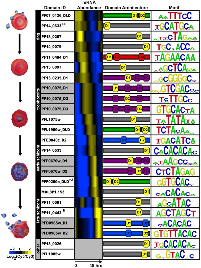
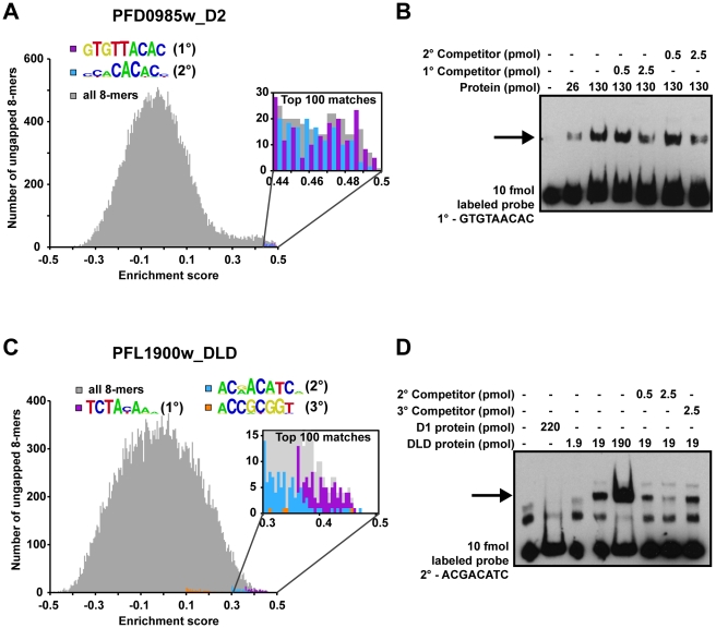
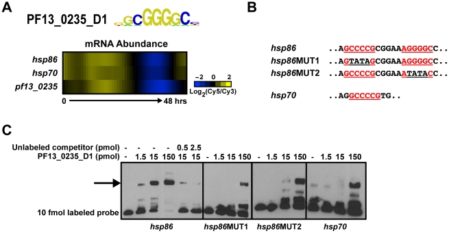
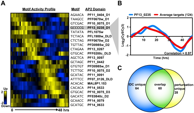
The molecular mechanisms underlying transcriptional regulation in apicomplexan parasites remain poorly understood. Recently, the Apicomplexan AP2 (ApiAP2) family of DNA binding proteins was identified as a major class of transcriptional regulators that are found across all Apicomplexa. To gain insight into the regulatory role of these proteins in the malaria parasite, we have comprehensively surveyed the DNA-binding specificities of all 27 members of the ApiAP2 protein family from Plasmodium falciparum revealing unique binding preferences for the majority of these DNA binding proteins. In addition to high affinity primary motif interactions, we also observe interactions with secondary motifs. The ability of a number of ApiAP2 proteins to bind multiple, distinct motifs significantly increases the potential complexity of the transcriptional regulatory networks governed by the ApiAP2 family. Using these newly identified sequence motifs, we infer the trans-factors associated with previously reported plasmodial cis-elements and provide evidence that ApiAP2 proteins modulate key regulatory decisions at all stages of parasite development. Our results offer a detailed view of ApiAP2 DNA binding specificity and take the first step toward inferring comprehensive gene regulatory networks for P. falciparum.


Conflict of interest statement

The authors have declared that no competing interests exist.

Figures

Figure 1
Figure 1. PBM derived motifs for P. falciparum ApiAP2 domains.
The first column lists the PlasmoDB gene ID and the corresponding AP2 domain tested on the PBM. D1, D2, and D3 refer to the AP2 domains numbered from the N- to C-terminus of each protein; and DLD indicates two domains and a short linker region. Gray shading in this column identifies different AP2 domains from the same protein. Column 2 depicts the relative mRNA abundance profiles for the ApiAP2 genes during the intraerythrocytic developmental cycle (IDC) . Genes that are not expressed during the IDC are in gray. The third column illustrates the number of domains, their architecture and approximate location in the sequence of each ApiAP2 protein (protein sizes are not to scale). The protein colours correspond to the number and arrangement of AP2 domains. Proteins with a single AP2 domain are in gray, two non-tandem AP2 domains are shaded blue, proteins with two tandem AP2 domains are shown in green, triple non-tandem domains are in purple, and the tandem double with an additional third domain is in red. The AP2 domain(s) that binds to the corresponding motif is depicted in yellow. The last column represents the highest scoring motif as a graphical representation of each position weight matrix and visualized using enoLOGOS . Motifs previously identified in the literature are marked as follows: * ; ; ; and § .
Figure 2
Figure 2. Secondary motifs represent both high- and low- affinity binding sites of ApiAP2 proteins.
A) A histogram for PFD0985w_D2 enrichment scores (E-scores) of all ungapped 8-mers (gray) demonstrates that both primary (purple bars) and secondary motifs (blue bars) are enriched in the tail area indicating high affinity interactions. A close-up view (inset) indicates that the top 100 8-mer matches for both motifs show a mixed distribution of E-scores, suggesting that PFD0985w_D2 will bind both motifs equally. B) EMSA competitions between PFD0985w_D2 primary and secondary motifs. The arrow denotes shifted probes. Competition with unlabeled probes of either the primary or secondary motif illustrate that both compete for binding equally well. No binding is observed in the absence of either motif (data not shown). The same experiment using labeled secondary motif probe yielded similar results (data not shown). Probes were labeled with biotin and all competitors are unlabeled. C) A histogram for PFL1900w_DLD E-scores of all ungapped 8-mers portrays similar enrichment of motifs at the high end of E-scores. A close-up view of the top 100 8-mers (inset) reveals that there is a clear preference for the primary motif (purple bars) over the secondary (blue bars), and the secondary motif over the tertiary (orange bars) suggesting different binding affinities. D) EMSA competitions of the PFL1900w_DLD secondary motif with unlabeled probes of the secondary and tertiary motifs, illustrate that the tertiary motif does not compete for binding with the secondary motif. No binding is observed with an unrelated oligonucleotide probe (data not shown). Shifted probes are indicated by the arrow.
Figure 3
Figure 3. PF13_0235_D1 binds the G-box and is a putative regulator of heat shock genes.
A) Transcript abundance profiles of pf13_0235, hsp86 and hsp70 genes portrays similar timing . B) Partial sequences of EMSA probes from the upstream sequences of hsp86 and hsp70. G-box motifs are underlined and shown in red. Mutations in the G-box sequence are underlined in black. C) EMSAs with probes from the upstream sequence of hsp86 and hsp70 illustrate the ability of PF13_0235_D1 to bind the target sequences. Binding to the hsp86 probe is mediated through both G-boxes as mutation of the first or second sequence significantly reduces binding. No binding is observed with an unrelated oligonucleotide (data not shown). The arrow denotes shifted probes. Probes were labeled with biotin and all competitors are unlabeled.
Figure 4
Figure 4. Activity profiles for AP2 motifs and refinement of target gene predictions during the IDC.
A) The heat map shows the motif activity profiles based on mutual information content between the motifs and genes expressed at each timepoint of the IDC. Target genes with expression profiles similar to the activity profiles and containing the motif of interest were identified. B) A plot of the average mRNA abundance for target genes with matches to the G-box motif in their upstream sequences is depicted. This is compared with the relative mRNA abundance data for pf13_0235, the ApiAP2 factor that binds to the G-box. There is a strong positive correlation (0.97) between pf13_0235 expression and the expression of its predicted targets. C) The Venn diagram shows the overlap between IDC co-expressed targets and perturbation co-expressed targets. Half of PF13_0235 targets are shared between both datasets, and include ribosomal and heat shock genes.
Figure 5
Figure 5. ApiAP2 motifs are conserved in var gene promoters.
Matches to ApiAP2 motifs were identified using position weight matrices for each motif. Searches were performed on 3 kb upstream of the ATG start codon or until the next upstream gene was encountered (shorter lines). The presence and position of motifs are conserved in the promoters of different types of var genes. The previously described SPE1 and SPE2 motifs in upsB var genes were found as well as occurrences of the PF11_0442 motif in all types of var genes.

References

    1. WHO. World Malaria Report 2009 2009.
    1. Bozdech Z, Llinás M, Pulliam BL, Wong ED, Zhu J, et al. The transcriptome of the intraerythrocytic developmental cycle of Plasmodium falciparum. PLoS Biol. 2003;1:E5. - PMC - PubMed
    1. Florens L, Washburn MP, Raine JD, Anthony RM, Grainger M, et al. A proteomic view of the Plasmodium falciparum life cycle. Nature. 2002;419:520–526. - PubMed
    1. Hall N, Karras M, Raine JD, Carlton JM, Kooij TW, et al. A comprehensive survey of the Plasmodium life cycle by genomic, transcriptomic, and proteomic analyses. Science. 2005;307:82–86. - PubMed
    1. Lasonder E, Ishihama Y, Andersen JS, Vermunt AM, Pain A, et al. Analysis of the Plasmodium falciparum proteome by high-accuracy mass spectrometry. Nature. 2002;419:537–542. - PubMed

Publication types

MeSH terms