. 2010 Oct 27;5(10):e13695.
doi: 10.1371/journal.pone.0013695.

Splice isoforms of the polyglutamine disease protein ataxin-3 exhibit similar enzymatic yet different aggregation properties

Affiliations

Splice isoforms of the polyglutamine disease protein ataxin-3 exhibit similar enzymatic yet different aggregation properties

Ginny Marie Harris et al. PLoS One. .

Abstract

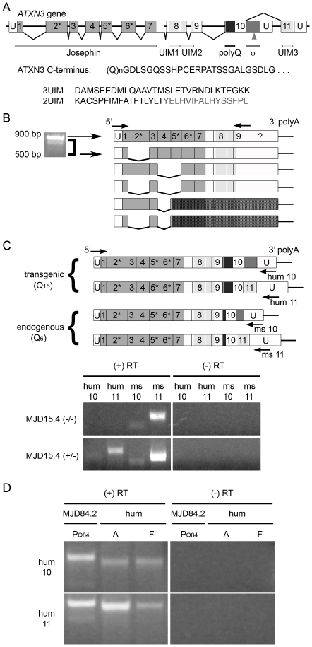
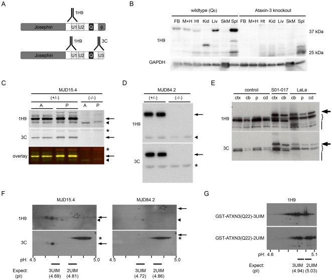
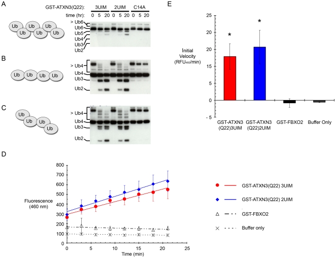
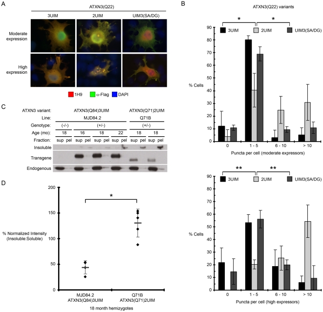
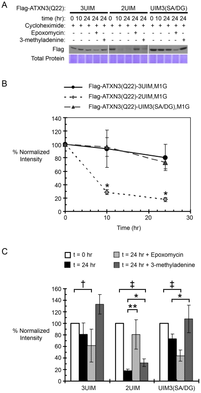
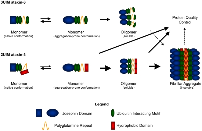
Protein context clearly influences neurotoxicity in polyglutamine diseases, but the contribution of alternative splicing to this phenomenon has rarely been investigated. Ataxin-3, a deubiquitinating enzyme and the disease protein in SCA3, is alternatively spliced to encode either a C-terminal hydrophobic stretch or a third ubiquitin interacting motif (termed 2UIM and 3UIM isoforms, respectively). In light of emerging insights into ataxin-3 function, we examined the significance of this splice variation. We confirmed neural expression of several minor 5' variants and both of the known 3' ataxin-3 splice variants. Regardless of polyglutamine expansion, 3UIM ataxin-3 is the predominant isoform in brain. Although 2UIM and 3UIM ataxin-3 display similar in vitro deubiquitinating activity, 2UIM ataxin-3 is more prone to aggregate and more rapidly degraded by the proteasome. Our data demonstrate how alternative splicing of sequences distinct from the trinucleotide repeat can alter properties of the encoded polyglutamine disease protein and thereby perhaps contribute to selective neurotoxicity.


Conflict of interest statement

Competing Interests: The authors have declared that no competing interests exist.

Figures

Figure 1
Figure 1. Ataxin-3 is alternatively spliced in ATXN3 YAC transgenic and human brain.
(A) Schematic representation of the ATXN3 gene showing exons that encode specific functional domains. Untranslated regions (U) are not drawn to scale. The splicing pattern of the originally identified 2UIM ataxin-3 transcript is shown below, while above is shown the alternative splicing that links exon 10 to exon 11, generating 3UIM ataxin-3. Asterisks indicate exons that encode amino acids comprising the catalytic triad, polyQ denotes the polyglutamine domain, and the arrowhead indicates a polymorphic Tyr/Stop-encoding residue within the hydrophobic domain (Φ) of the C-terminus of 2UIM ataxin-3. C-terminal amino acid sequences are shown below the diagram, beginning with shared sequence in both isoforms extending from the polyQ domain, followed by the divergent sequences for the 2UIM and 3UIM isoforms; residues omitted in some SNP variants of the 2UIM isoform are shown in grey. (B) Diagram showing 5′ ataxin-3 splice variants identified and confirmed by sequencing. Multiple variants are detectable in mature mRNA from adult murine brain (and fetal brain, data not shown) by RT-PCR, using primers targeting the 5′UTR/exon 1 junction and exon 9 (arrows). All identified splice variants that maintain the open reading frame eliminate at least one catalytic triad residue, and thus are not likely to encode an active DUB. Darkly shaded areas are downstream of a frameshift-induced stop codon. (C–D) Endogenous Atxn3 and transgenic ATXN3 “full length” splice variants were amplified by RT-PCR using species-specific (human, hum; murine, ms) and sequence-specific (10-exon 2UIM-encoding, 10; 11-exon 3UIM-encoding, 11) primers. 10-exon and 11-exon variants are both detectable in mature mRNA: (C) endogenous Atxn3 from all murine samples and unexpanded ATXN3 from MJD15.4(+/−) brain; and (D) expanded ATXN3 from MJD84.2(+/−) brain, and unexpanded ATXN3 from pooled human brain tissue (hum). Perinatal day 1–3 (PQ84), adult (A), or fetal (F) sources were used, as indicated. Note: Primers are not drawn to scale; see Materials and Methods for exact sequences and locations.
Figure 2
Figure 2. 3UIM ataxin-3 is the predominant protein isoform in murine and human brain tissue.
(A) Diagram of 2UIM (upper) and 3UIM (lower) ataxin-3 variants showing recognition sites for 1H9 mAb, which recognizes both isoforms, and α-ataxin-3C, which recognizes only 3UIM ataxin-3. (B) Western blot of wildtype (Q6) or Atxn3 knockout mouse tissue lysates, probed for endogenous murine ataxin-3 (1H9) and GAPDH. Tissues include forebrain (FB), midbrain and hindbrain (M+H), heart (Ht), kidney (Kid), liver (Liv), skeletal muscle (SkM), and spleen (Spl). Although various putative tissue-specific splice isoforms exist, there is only one predominant isoform in brain tissue. (C–D) The major isoform (arrow) of human ataxin-3 is recognized by the 3UIM-specific antibody (3C) and 1H9 in brain tissue from hemizygous transgenic (+/−) MJD15.4 (C, Q15) and MJD84.2 mice (D, Q84), whereas endogenous Q6 ataxin-3 (arrowhead) is recognized only by 1H9 in hemizygous transgenic mice and wildtype (−/−) controls. Perinatal day 1–3 (P), adult (A), and non-specific 3C signal (*), as shown. (E) Both 1H9 and 3C recognize the predominant non-expanded ataxin-3 isoforms (brackets) in healthy controls and SCA3 patients (S01–017 and LaLa), as well as the predominant expanded isoform in SCA3 patients (bold arrows). Lower molecular weight bands (bars) are preferentially recognized by 1H9; cortex (ctx), cerebellum (cb), putamen (p), caudate (cd) sources, as indicated. (F,G) 2D-Western blot analysis was used to distinguish 2UIM from 3UIM ataxin-3 protein. IPG range and predicted isoelectric points (pI) of each isoform are shown. (F) In brain lysates of MJD15.4 (Q15)or MJD84.2 (Q84) YAC transgenic mice, 1H9 recognizes multiple species including endogenous murine (arrowhead) and 3UIM ataxin-3 transprotein (arrow), but does not detect any 2UIM ataxin-3 (which would be 1H9-positive, 3C-negative, 0.5 kDa larger than 3UIM ataxin-3, with a basic shift in pI, as indicated by the open arrow). The prominent band detected by 3C (*) is nonspecific. (G) 2D-Western of 50 ng purified recombinant GST-tagged ataxin-3 isoforms shows that unexpanded Q22 2UIM GST-ataxin-3 is detected as readily as 3UIM GST-ataxin-3.
Figure 3
Figure 3. 2UIM and 3UIM ataxin-3 display similar DUB activity against defined ubiquitin chains in vitro.
(A–C) Recombinant GST-ATXN3(Q22) (3UIM or 2UIM) can cleave K48-linked hexaubiquitin (A), K63-linked tetraubiquitin (B), and mixed linkage K48-K63-K48 tetraubiquitin (C) chains. Results with catalytically inactive GST-ataxin-3 (C14A mutant) are also shown. (D–E) Recombinant 2UIM and 3UIM GST-ATXN3(Q22) cleave Ub-AMC at a similar rate. (D) Ub-AMC reaction curves. Both 3UIM and 2UIM ataxin-3 area able to cleave Ub-AMC, while reactions with either an unrelated control protein (the non-DUB F-box protein FBXO2) or buffer only show no cleavage. Error bars show standard deviations. (E) There is no significant difference between the initial reaction velocity of 2UIM and 3UIM ataxin-3(Q22) (p>0.4 by a 2 tailed heteroscedastic Student's t-test).
Figure 4
Figure 4. 2UIM ataxin-3 is more prone to aggregate than 3UIM ataxin-3.
(A) Representative immunofluorescence of Cos7 cells transiently expressing Flag-tagged ataxin-3(Q22) splice isoforms or the UIM3(SA/DG) mutant. Cells were gated by fluorescence intensity into populations of moderate and high expressors. (B) Quantification of puncta per cell in (A). Error bars represent the standard deviation within each bin. Frequency distributions differ significantly between ATXN3(Q22)2UIM and ATXN3(Q22)3UIM and between ATXN3(Q22)2UIM and ATXN3(Q22)UIM3(SA/DG) mutant ataxin-3 (*p<0.0001, ** p<1×10-11), but not between ATXN3(Q22)3UIM and ATXN3(Q22)UIM3(SA/DG) mutant ataxin-3 by a χ2 test for independence, df  = 3. (C) Supernatant (sup) and pellet (pel) fractions of non-denaturing RIPA brain lysates from aged MJD84.2 (ATXN3(Q84)3UIM) and Q71B (ATXN3(Q71)2UIM) hemizygous transgenic mice were analyzed by Western blot with 1H9 anti-ataxin-3 antibody. Insoluble microaggregates are detected at the base of lane wells, whereas soluble transprotein and endogenous ataxin-3 are visualized within the resolving gel. (D) Quantification of the ratio of insoluble to soluble ataxin-3 transprotein seen in (C). 3UIM-predominant MJD84.2 mice show a significantly lower ratio of insoluble:soluble transprotein than 2UIM-only Q71B mice (*p<0.0005 by a 1 tailed heteroscedastic Student's t-test).
Figure 5
Figure 5. 2UIM ataxin-3 is a less stable protein than 3UIM ataxin-3 and is subject to rapid proteasomal degradation.
(A) Representative cycloheximide “pulse-chase” in Cos7 cells transiently transfected with Flag-tagged ataxin-3(Q22) constructs; ataxin-3 levels are visualized by anti-Flag Western blotting and total protein levels are visualized by Coomassie Brilliant Blue staining of the PVDF membrane. (B) Quantification of ataxin-3 levels during a 24 hour incubation with cycloheximide: ATXN3(Q22)2UIM is degraded significantly faster than ATXN3(Q22)3UIM or ATXN3(Q22)UIM3(SA/DG) mutant ataxin-3 at 10 and 24 hours (*p<0.02 by a two-tailed heteroscedastic Student's t-test). (C) Quantification of ataxin-3 levels during a 24 hour cycloheximide incubation in the absence or presence of the proteasomal inhibitor epoxomycin or the macroautophagy inhibitor 3-methyladenine; loss of protein at 24 hours is rescued by proteasomal inhibition for ATXN3(Q22)2UIM and by inhibition of macroautophagy for ATXN3(Q22)3UIM and ATXN3(Q22)UIM3(SA/DG) mutant ataxin-3 (†p<0.05 or ‡p<0.01 compared to time zero; *p<0.05 or **p<0.01 compared to 24 hour time point by paired one-tailed Student's t-tests). In B and C, densitometry analyses are plotted as the percentage of signal at time zero, normalized to total protein signal.
Figure 6
Figure 6. Model for the differential aggregation properties and processing of 2UIM and 3UIM ataxin-3.
In the absence of polyglutamine expansion, 3UIM ataxin-3 follows a multi-domain aggregation mechanism to generate limited oligomeric species without detectable formation of SDS-insoluble fibrillar aggregates. In contrast, 2UIM ataxin-3 exists in at least two monomeric states: the native conformation, in which the hydrophobic tail remains buried and protected from the aqueous environment, and an aggregation-prone conformation with an exposed hydrophobic tail. The aggregation prone monomer can revert to the native conformation or oligomerize through both the self-association propensity of the Josephin domain (like 3UIM ataxin-3) and hydrophobic interactions of the 2UIM-specific domain. Within 2UIM oligomers, the hydrophobic C-termini will associate, increasing the local polyglutamine concentration beyond that seen in 3UIM oligomers, favoring formation of detergent-insoluble aggregates. Unstable forms of monomer and oligomer are subject to protein quality control mechanisms, including proteasomal degradation for 2UIM ataxin-3. Insoluble fibrils, which are less well handled by protein quality control systems, accumulate as biochemically and microscopically detectable aggregates.

Similar articles

Cited by

References

    1. Orr HT, Zoghbi HY. Trinucleotide repeat disorders. Annu Rev Neurosci. 2007;30:575–621. - PubMed
    1. Johnson JM, Castle J, Garrett-Engele P, Kan Z, Loerch PM, et al. Genome-wide survey of human alternative pre-mRNA splicing with exon junction microarrays. Science. 2003;302(5653):2141–4. - PubMed
    1. Wang ET, Sandberg R, Luo S, Khrebtukova I, Zhang L, et al. Alternative isoform regulation in human tissue transcriptomes. Nature. 2008;456(7221):470–6. - PMC - PubMed
    1. Tsunemi T, Ishikawa K, Jin H, Mizusawa H. Cell-type-specific alternative splicing in spinocerebellar ataxia type 6. Neuroscience Letters. 2008;447(1):78–81. - PubMed
    1. Tadokoro K, Yamazaki-Inoue M, Tachibana M, Fujishiro M, Nagao K, et al. Frequent occurrence of protein isoforms with or without a single amino acid residue by subtle alternative splicing: the case of Gln in DRPLA affects subcellular localization of the products. J Hum Genet. 2005;50(8):382–94. - PubMed

Publication types

LinkOut - more resources