

Effects of Ginkgo biloba on cerebral blood flow assessed by quantitative MR perfusion imaging: a pilot study
- PMID: 21061003
- PMCID: PMC3163160
- DOI: 10.1007/s00234-010-0790-6
Effects of Ginkgo biloba on cerebral blood flow assessed by quantitative MR perfusion imaging: a pilot study
Abstract
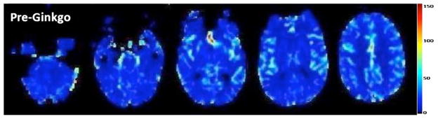
Introduction: Extract of Ginkgo biloba (EGb), a dietary supplement used for a number of conditions including dementia, has been suggested to increase cerebral bloodflow (CBF). The purpose of this study was to determine if changes in CBF could be detected by dynamic susceptibility contrast-enhanced magnetic resonance imaging (DSC-MRI)in elderly human subjects taking EGb.
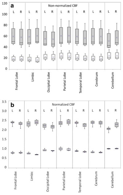
Methods: DSC-MRI was performed in nine healthy men(mean age 61±10 years) before and after 4 weeks of 60 mg EGb taken twice daily. One subject underwent six consecutive scans to evaluate intrasubject reproducibility. CBF values were computed before and after EGb, and analyzed at three different levels of spatial resolution, using voxel-based statistical parametric mapping (SPM), and regions of interest in different lobes, and all regions combined.
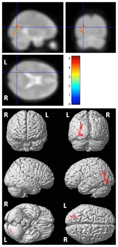
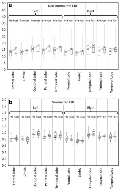
Results: Normalized intrasubject CBF (nCBF) measurements had a standard deviation of 7% and 4% in gray and white matter (WM) regions, respectively. SPM using an uncorrected, voxel-level threshold of P≤0.001 showed a small CBF increase in the left parietal-occipital region.CBF in individual lobar regions did not show any significant change post-EGb, but all regions combined showed a significant increase of non-normalized CBF after EGb (15% in white and 13% in gray matter, respectively, P≤0.0001).
Conclusion: nCBF measured by DSC-MRI has good intrasubject reproducibility. In this small cohort of normal elderly individuals, a mild increase in CBF is found in the left parietal-occipital WM after EGb, as well as a small but statistically significant increase in global CBF.
Conflict of interest statement
Figures
References
-
- Schneider LS. Ginkgo biloba extract and preventing Alzheimer disease. JAMA. 2008;300:2306–2308. - PubMed
-
- Sun BL, Yuan H, Xia YMF, ZLZSM, Wang LX. Effects of extract of Ginkgo biloba on intracranial pressure, cerebral perfusion pressure, and cerebral blood flow in a rat model of subarachnoid haemorrhage. Int J Neurosci. 2007;117:655–665. - PubMed
-
- Sun BL, Zhang J, Xia WXC, ZLYMF, Zhang SM, Ye WJ, Yuan H. Effects of extract of Ginkgo biloba on spasms of the basilar artery and cerebral microcirculatory perfusion in rats with subarachnoid hemorrhage. Clin Hemorheol Microcirc. 2003;29:231–238. - PubMed
Publication types
MeSH terms
Substances
Grants and funding
LinkOut - more resources
Full Text Sources
