. 2011 Feb;42(1):3-13.
doi: 10.1007/s10735-010-9302-6.

Characterization of basal pseudopod-like processes in ileal and colonic PYY cells

Affiliations

Characterization of basal pseudopod-like processes in ileal and colonic PYY cells

Diego V Bohórquez et al. J Mol Histol. 2011 Feb.

Abstract

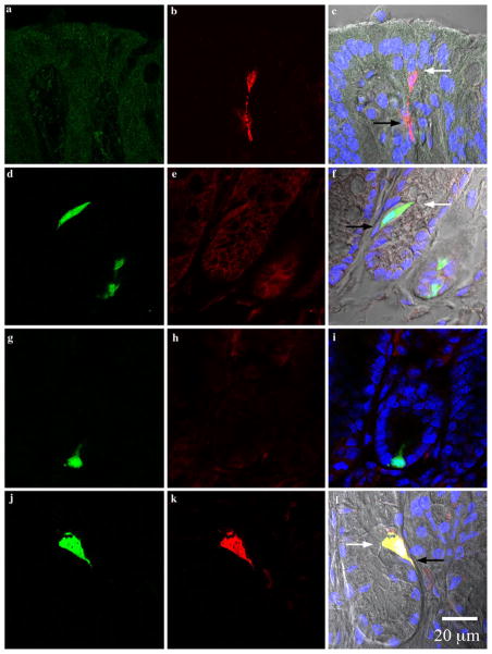
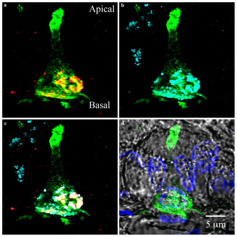
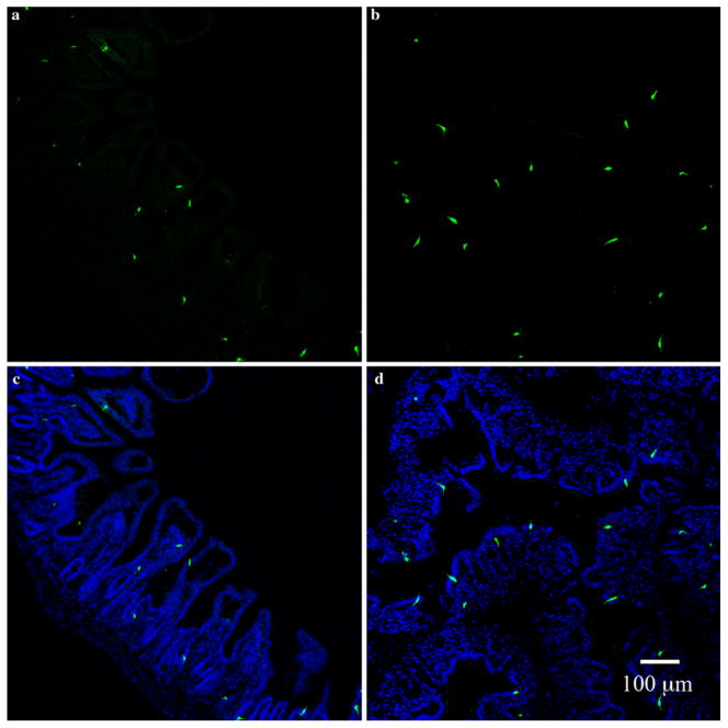
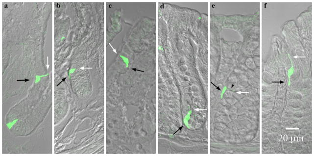
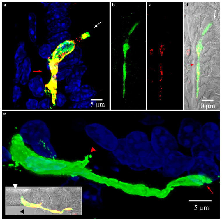
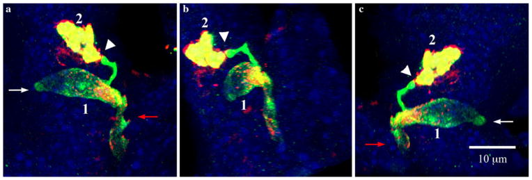
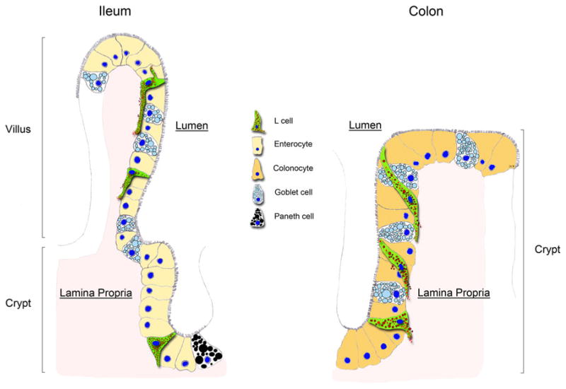
The peptide tyrosine tyrosine (PYY) is produced and secreted from L cells of the gastrointestinal mucosa. To study the anatomy and function of PYY-secreting L cells, we developed a transgenic PYY-green fluorescent protein mouse model. PYY-containing cells exhibited green fluorescence under UV light and were immunoreactive to antibodies against PYY and GLP-1 (glucagon-like peptide-1, an incretin hormone also secreted by L cells). PYY-GFP cells from 15 μm thick sections were imaged using confocal laser scanning microscopy and three-dimensionally (3D) reconstructed. Results revealed unique details of the anatomical differences between ileal and colonic PYY-GFP cells. In ileal villi, the apical portion of PYY cells makes minimal contact with the lumen of the gut. Long pseudopod-like basal processes extend from these cells and form an interface between the mucosal epithelium and the lamina propria. Some basal processes are up to 50 μm in length. Multiple processes can be seen protruding from one cell and these often have a terminus resembling a synapse that appears to interact with neighboring cells. In colonic crypts, PYY-GFP cells adopt a spindle-like shape and weave in between epithelial cells, while maintaining contact with the lumen and lamina propria. In both tissues, cytoplasmic granules containing the hormones PYY and GLP-1 are confined to the base of the cell, often filling the basal process. The anatomical arrangement of these structures suggests a dual function as a dock for receptors to survey absorbed nutrients and as a launching platform for hormone secretion in a paracrine fashion.


Figures

Fig. 1
Fig. 1
Photomicrographs from colonic crypts. All sections are aligned vertically with the lumen on top. White and black arrows point to the lumen and lamina propria, respectively. Colors represent the following: green green fluorescent protein (GFP), red PYY, blue DAPI, and gray differential interference contrast (DIC) and are merged from the left and middle columns. ac Sections from wild type Swiss Webster mice stained for GFP (a) and PYY (b). GFP is absent and PYY cells appeared to have a spindle-like shape in the colon of wild type mice. df Sections from PYY-GFP mice incubated with secondary antibodies alone (DyLight488-conjugated donkey anti-chicken and Cy3-conjugated donkey anti-rabbit). Colonic cells from transgenic PYY-GFP mice expressed endogenous GFP and non-specific binding of secondary antibodies was absent. gi Sections from a PYY-GFP mouse incubated in the presence of chicken anti-GFP and rabbit anti-PYY pre-adsorbed in excess (10 μM) of PYY peptide. The specificity of the antibody to PYY is shown by the absence of immunoreactivy after pre-absorption with synthetic PYY. jl PYY-GFP sections incubated in the presence of chicken anti-GFP and rabbit anti-PYY antibodies. PYY immunoreactivity with the antibody exclusively co-localized with GFP positive cells (l, yellow). Please refer to online version of the article for colored figures
Fig. 2
Fig. 2
Colocalization of PYY and glucagon-like peptide 1 (GLP-1) in PYY-GFP cells of the ileum. a PYY staining (red) reveals confinement of granules to the basal end of the PYY-GFP cell. b GLP-1 staining (cyan) shows colocalization at the basal side of the PYY-GFP cell. c Image overlay of PYY and GLP-1 granules colocalized at the base of PYY-GFP cell (green GFP, red PYY, cyan GLP-1 and white overlapping of PYY and GLP-1. Staining for PYY (red) and GLP-1 (cyan) are projected as maximal intensities. Note that not all PYY and GLP-1 staining colocalizes within the same granules. This has been previously demonstrated by electron immu-nogold staining (Nilsson et al. 1991). d Position of PYY-GFP cell with respect to other cells of the epithelium (green GFP, blue DAPI and gray DIC)
Fig. 3
Fig. 3
Distribution of PYY-GFP cells in the ileum and colon. Sections of ileum (a, c) and colon (b, d) from a PYY-GFP mouse stained with chicken anti-GFP (a, b) and composite with DAPI nuclei (blue) stain (c, d). PYY-GFP cells appear to be more abundant in the colon than in the ileum. Note the spindle-like anatomy of PYY-GFP cells in the colon compared to the L shape of those in the ileum
Fig. 4
Fig. 4
PYY-GFP cell morphology in the ileum versus colon. White and black arrows indicate the lumen and lamina propria respectively. ac In the ileum a cells were columnar or flask-shaped at the bottom of the crypts and pseudopod-like basal processes were absent. Further up the crypt (b) towards the villus tip (c) basal processes became evident. df In the colon, flask-shaped cells were evident at the bottom of the crypts (d). A neck-like projection (black arrow head) and a basal process were evident in the cell in the middle of the crypt (e). Towards the apex of the crypt, this neck-like projection extended to maintain contact with the lumen, while the basal process remained in contact with the lamina propria (f). Note the sigmoidal appearance of the cell to maintain contact with the lumen as well as the lamina propria
Fig. 5
Fig. 5
Pseudopod-like basal processes in PYY-GFP cells of the ileum. White and red arrows indicate the lumen and lamina propria respectively. Sections imaged were 15 μm thick. a Maximal projection view of typical PYY-GFP cell in a villus of the ileum. The pseudopod-like process runs on the basal side, in between the lamina propria and the base of other epithelial cells. The basal process typically extends downward towards the bottom of the crypt. bd PYY-GFP cell with a long basal process extending along the base of adjacent epithelial cells. The total length of the cell is greater than 70 μm and the basal process is approximately 50 μm long. b Green GFP, c red PYY, d composite with DIC (gray). e Maximal projection view of PYY-GFP cell in a villus from the ileum. Inset shows the cell position with respect to the brush border (white arrow head) and lamina propria (black arrow head) (green GFP, red PYY staining, yellow merged images, gray DIC). The pseudopod-like basal process often ended in a bifurcation (red arrow). There are some smaller projections (red arrow head) that extended from the body of the cell
Fig. 6
Fig. 6
Relationship of a PYY-GFP cell with another PYY-GFP cell through synapse-like structures at the end of pseudopod-like processes. White and red arrows indicate the lumen and lamina propria respectively. Section imaged was 15 μm thick. ac Views of three-dimensionally reconstructed PYY-GFP cells in a villus of the ileum from different angles. The synapse-like projection at the end of pseudopod-like process from cell 1 that makes contact with cell 2 is shown by the white arrowhead. Green GFP, red PYY staining, yellow merged images, blue DAPI
Fig. 7
Fig. 7
Representation of PYY cells in the ileum versus colon. In the ileum, PYY cells (L cells) are flask-shaped at the crypt whereas, in the villus, a long pseudopod-like process extends at their base. The basal processes usually extend toward the crypt. Occasionally, more than one process can extend from the body of the cells toward other epithelial cells. In the colon, PYY cells are flask-shaped at the bottom of the crypt. However, at the apex of the crypt, PYY cells weave through other epithelial cells and the tip of the cell runs along the crypt’s lumen. The base of the same PYY cell runs vertically along the crypt’s lamina propria. Whereas in the ileum PYY cells maintain minimal contact with the gut lumen, in the colon PYY cells appear to have a larger surface area exposed to the lumen

References

    1. Alumets J, Ekelund M, Munshid H, HÂkanson R, Loren I, Sundler F. Topography of somatostatin cells in the stomach of the rat: possible functional significance. Cell Tiss Res. 1979;202:177–188. - PubMed
    1. Batterham R, Cowley M, Small C, Herzog H, Cohen M, Dakin C, Wren A, Brynes A, Low M, Ghatei M. Gut hormone PYY3–36 physiologically inhibits food intake. Nature. 2002;418:650–654. - PubMed
    1. Batterham R, Heffron H, Kapoor S, Chivers J, Chandarana K, Herzog H, Le Roux C, Thomas E, Bell J, Withers D. Critical role for peptide YY in protein-mediated satiation and body-weight regulation. Cell Metabol. 2006;4:223–233. - PubMed
    1. Chandra R, Samsa L, Vigna S, Liddle R. Pseudopod-like basal cell processes in intestinal cholecystokinin cells. Cell Tiss Res. 2010 doi: 10.1007/s00441-010-0997-1. - DOI - PMC - PubMed
    1. Cheng H, Leblond CP. Origin, differentiation and renewal of the four main epithelial cell types in the mouse small intestine III. Entero-endocrine cells. Am J Anat. 1974;141:503–519. - PubMed

Publication types

LinkOut - more resources