Contribution of perfusion-weighted magnetic resonance imaging in the differentiation of meningiomas and other extra-axial tumors: case reports and literature review
- PMID: 21061142
- PMCID: PMC3116130
- DOI: 10.1007/s11060-010-0445-9
Contribution of perfusion-weighted magnetic resonance imaging in the differentiation of meningiomas and other extra-axial tumors: case reports and literature review
Abstract
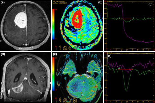
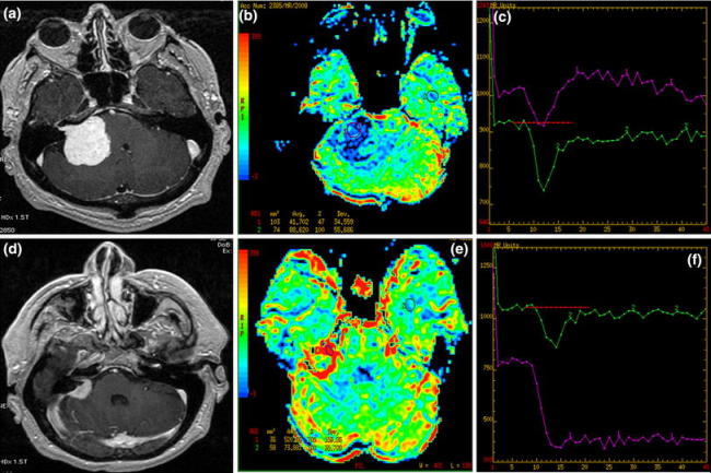
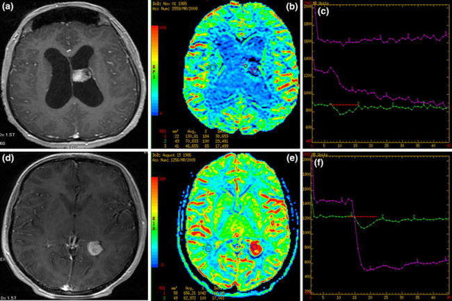
We present six cases of extra-axial lesions: three meningiomas [including one intraventricular and one cerebellopontine angle (CPA) meningioma], one dural metastasis, one CPA schwannoma and one choroid plexus papilloma which were chosen from a larger cohort of extra-axial tumors evaluated in our institution. Apart from conventional MR examinations, all the patients also underwent perfusion-weighted imaging (PWI) using dynamic susceptibility contrast method on a 1.5 T MR unit (contrast: 0.3 mmol/kg, rate 5 ml/s). Though the presented tumors showed very similar appearance on conventional MR images, they differed significantly in perfusion examinations. The article draws special attention to the usefulness of PWI in the differentiation of various extra-axial tumors and its contribution in reaching final correct diagnoses. Finding a dural lesion with low perfusion parameters strongly argues against the diagnosis of meningioma and should raise a suspicion of a dural metastasis. In cases of CPA tumors, a lesion with low relative cerebral blood volume values should be suspected to be schwannoma, allowing exclusion of meningioma to be made. In intraventricular tumors arising from choroid plexus, low perfusion parameters can exclude a diagnosis of meningioma. In our opinion, PWI as an easy and quick to perform functional technique should be incorporated into the MR protocol of all intracranial tumors including extra-axial neoplasms.
Figures


Similar articles
-
Imaging of cerebellopontine angle lesions: an update. Part 2: intra-axial lesions, skull base lesions that may invade the CPA region, and non-enhancing extra-axial lesions.Eur Radiol. 2007 Nov;17(11):2908-20. doi: 10.1007/s00330-007-0680-4. Epub 2007 Jun 14. Eur Radiol. 2007. PMID: 17569053 Review.
-
Contribution of dynamic contrast MR imaging to the differentiation between dural metastasis and meningioma.Neuroradiology. 2004 Aug;46(8):642-8. doi: 10.1007/s00234-004-1194-2. Epub 2004 Jun 30. Neuroradiology. 2004. PMID: 15232661 Clinical Trial.
-
Usefulness of PRESTO magnetic resonance imaging for the differentiation of schwannoma and meningioma in the cerebellopontine angle.Neurol Med Chir (Tokyo). 2013;53(7):482-9. doi: 10.2176/nmc.53.482. Neurol Med Chir (Tokyo). 2013. PMID: 23883559
-
Beyond the expected: a supratentorial ependymoma imitating a meningioma.Childs Nerv Syst. 2025 Mar 26;41(1):141. doi: 10.1007/s00381-025-06803-7. Childs Nerv Syst. 2025. PMID: 40133725
-
Association of an intracranial arteriovenous malformation and a meningioma.Neuroradiology. 1998 Sep;40(9):574-6. doi: 10.1007/s002340050646. Neuroradiology. 1998. PMID: 9808313 Review.
Cited by
-
Choroid Plexus Neoplasms: Toward a Distinction between Carcinoma and Papilloma Using Arterial Spin-Labeling.AJNR Am J Neuroradiol. 2015 Sep;36(9):1786-90. doi: 10.3174/ajnr.A4332. Epub 2015 May 28. AJNR Am J Neuroradiol. 2015. PMID: 26021621 Free PMC article.
-
Contribution of advanced neuroimaging in diagnosis of cerebral syphilitic gumma: a case report.Front Neurosci. 2024 Aug 14;18:1442176. doi: 10.3389/fnins.2024.1442176. eCollection 2024. Front Neurosci. 2024. PMID: 39206117 Free PMC article.
-
Measurement of choroid plexus perfusion using dynamic susceptibility MR imaging: capillary permeability and age-related changes.Neuroradiology. 2013 Dec;55(12):1447-54. doi: 10.1007/s00234-013-1290-2. Epub 2013 Oct 23. Neuroradiology. 2013. PMID: 24150596
-
A semi-automated volumetric software for segmentation and perfusion parameter quantification of brain tumors using 320-row multidetector computed tomography: a validation study.Neuroradiology. 2017 May;59(5):461-469. doi: 10.1007/s00234-017-1790-6. Epub 2017 Mar 24. Neuroradiology. 2017. PMID: 28341992
-
Central neurocytoma-positive and differential diagnosis: An example through a case report.SAGE Open Med Case Rep. 2023 Apr 7;11:2050313X231164280. doi: 10.1177/2050313X231164280. eCollection 2023. SAGE Open Med Case Rep. 2023. PMID: 37056701 Free PMC article.
References
Publication types
MeSH terms
LinkOut - more resources
Full Text Sources