. 2010 Nov;40(11):3043-53.
doi: 10.1002/eji.201040577. Epub 2010 Oct 27.

Apoptosis regulators Fas and Bim synergistically control T-lymphocyte homeostatic proliferation

Affiliations

Apoptosis regulators Fas and Bim synergistically control T-lymphocyte homeostatic proliferation

Karen A Fortner et al. Eur J Immunol. 2010 Nov.

Abstract

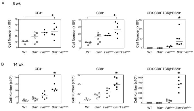
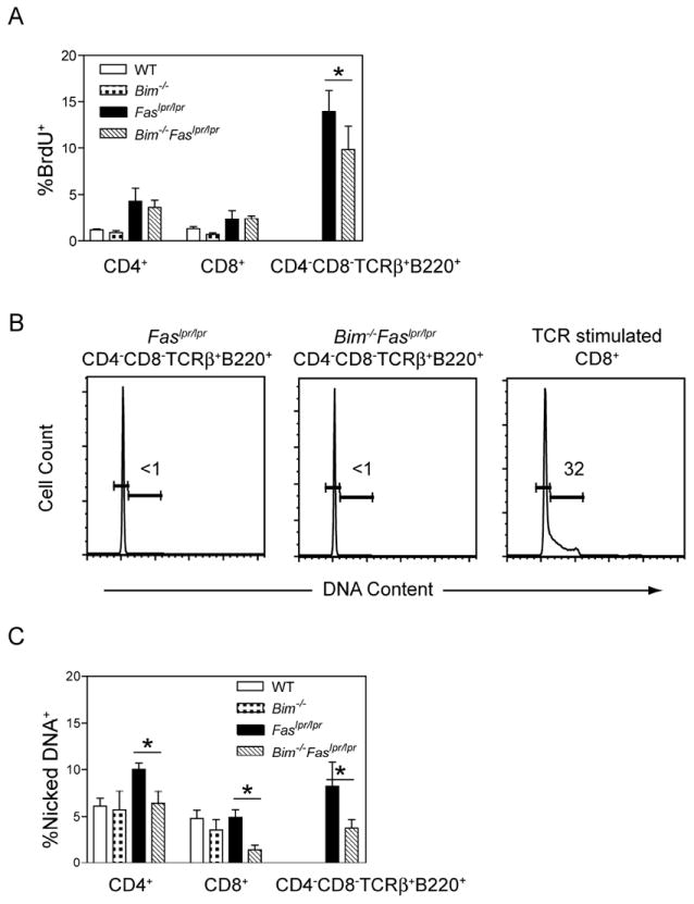
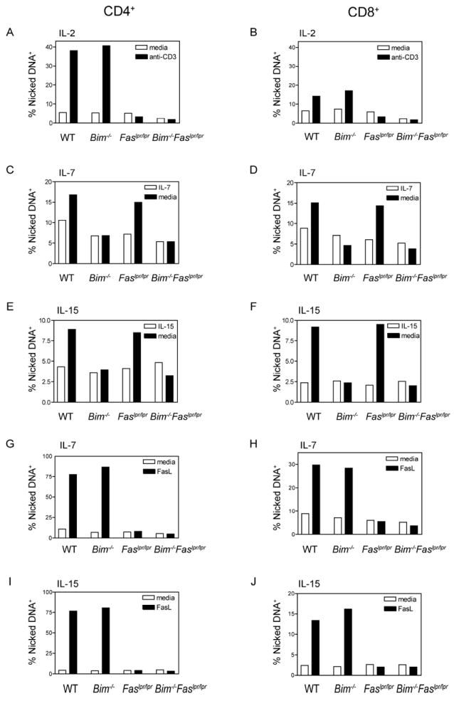
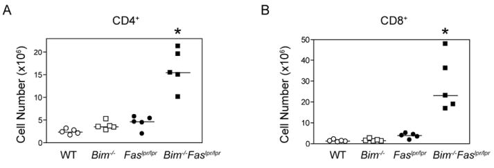
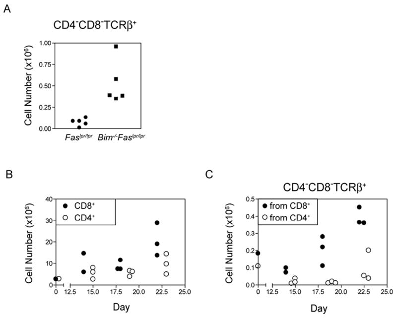
The size of the peripheral T-lymphocyte compartment is governed by complex homeostatic mechanisms that balance T-cell proliferation and death. Proliferation and survival signals are mediated in part by recurrent self-peptide/MHC-TCR interactions and signaling by the common γ chain-containing cytokine receptors, including those for IL-7 and IL-15. We have previously shown that the death receptor Fas (CD95/APO-1) regulates apoptosis in response to repeated TCR stimulation, whereas the Bcl-2 homology domain 3-only protein Bim mediates cytokine withdrawal-induced apoptosis. We therefore reasoned that these two molecules might cooperate in the regulation of homeostatic proliferation. In this study, we observe that the combined loss of Fas and Bim synergistically enhances the accumulation of T cells in lymphopenic host mice, and this is particularly pronounced for the unusual CD4(-) CD8(-) TCRαβ(+) T cells that are characteristic of Fas-deficient (Fas(lpr/lpr) ) mice. Our findings demonstrate that these CD4(-) CD8(-) TCRαβ(+) T cells arise from homeostatic proliferation of CD8(+) T cells. These studies also underscore the profound rate of baseline T-cell proliferation that likely occurs in wild-type mice even in the absence of foreign antigen, and the consequent need for its coordinated regulation by multiple death-signaling pathways.


Conflict of interest statement

The authors have no conflicting financial or commercial interests.

Figures

Figure 1
Figure 1. Accelerated lymphadenopathy in Bim-/-Faslpr/lpr mice
The absolute numbers of CD8+, CD4+, and CD4CD8TCRαβ+B220+ lymph node T cells recovered from wild-type (WT), Bim-/-, Faslpr/lpr, and Bim-/-Faslpr/lpr mice at (A) 8 wk and (B) 14 wk of age. Lymph node cells were stained for CD4, CD8, TCRβ, and CD45R (B220) and analyzed by flow cytometry. Cell numbers were calculated based on the percent positive cells. Each symbol represents an individual mouse analyzed (n=4-6 per group). Horizontal lines indicate the median number for each T cell subset. At 8 wk, differences in cell number were significant between Bim-/-Faslpr/lpr and wild-type T cells for both CD4+ and CD8+ T cells (ANOVA, Tukey post-test, *p< 0.05). At 14 wk, differences in cell number between Bim-/-Faslpr/lpr and the parental genotype T cells were significant for both CD4+ and CD8+ T cells (ANOVA, Tukey post-test, *p< 0.05). Differences in the number of CD4CD8TCRαβ+B220+ T cells between Bim-/-Faslpr/lpr and Faslpr/lpr were significant at both 8 and 14 wk (t test, *p<0.05).
Figure 2
Figure 2. Bim-/-Faslpr/lpr T cells manifest reduced apoptosis but equivalent rates of BrdU incorporation as compared with Faslpr/lpr T cells
(A) Age and sex-matched wild-type (WT), Bim-/-, Faslpr/lpr, and Bim-/-Faslpr/lpr mice received four intraperitoneal injections of BrdU (1 mg each) over 24 h. Lymph node cells were stained for surface expression of CD4, CD8, CD45R (B220), and analyzed for BrdU incorporation by flow cytometry. Shown are the mean and standard deviation for the percentage of BrdU+ cells in each T cell subset from 14 wk old mice (n=4 per strain). The percentages of BrdU+ cells between Bim-/-Faslpr/lpr and Faslpr/lpr T cells were statistically different for the CD4CD8TCRαβ+B220+, but not for the CD4+ and CD8+ T cell subsets (ANOVA, Tukey post-test, *p<0.05). These data are representative of two experiments. (B) Lymph node cells from age and sex-matched Faslpr/lpr and Bim-/-Faslpr/lpr mice were stained for surface expression of CD4, CD8, CD45R (B220), and TCRβ and analyzed for DNA content by Hoechst staining. Shown are representative Hoechst profiles for CD4CD8TCRαβ+B220+ T cells from Faslpr/lpr (n=3) and Bim-/-Faslpr/lpr mice (n=3) and for wild-type CD8+ T cells on day 3 after in vitro stimulation with plate-bound anti-CD3 and anti-CD28 antibodies. Number inserts are the percentage of cells in S/G2/M. (C) Single cell suspensions of lymph node cells from age and sex-matched wild-type (WT), Bim-/-, Faslpr/lpr, and Bim-/-Faslpr/lpr mice were cultured at 37°C for 4 h. The cells were then stained for surface expression of CD4, CD8, CD45R (B220), and TCRβ and analyzed for the presence of nicked DNA by TUNEL assay. The fraction of cells containing nicked DNA was calculated for each T cell subset. Shown are the mean and standard deviation of the percentage of apoptotic cells in each T cell subset from 14 wk old mice (n=3-4 per strain). The percentages of apoptotic cells between Bim-/-Faslpr/lpr and Faslpr/lpr T cells were statistically different for the CD4+, CD8+, and CD4CD8TCRαβ+B220+ T cell subsets (ANOVA, Tukey post-test, *p<0.05). These data are representative of two experiments.
Figure 3
Figure 3. Fas-deficient T cells are protected from CD3-restimulation-induced cell death whereas Bim-deficient T cells are protected from cytokine withdrawal
Single cell suspensions of lymph node cells from age and sex-matched wild-type (WT), Bim-/-, Faslpr/lpr, and Bim-/-Faslpr/lpr mice were stimulated with plate-bound anti-CD3 and anti-CD28 antibodies in the presence of (A, B) IL-2, (C, D, G, H) IL-7, or (E, F, I, J) IL-15. On day 2 of culture, the cells were removed from the anti-CD3/anti-CD28 antibody stimulation, washed, and cultured in the presence of IL-2, IL-7, or IL-15 consistent with the initial culture conditions. (A, B) On day 5, IL-2-cultured T cell blasts were restimulated with plate-bound anti-CD3 antibody for 6 h or left unstimulated. (C, D, E, F) T cells grown in either IL-7 or IL-15 for 5 days were washed three times to remove cytokines and then cultured in medium containing IL-7 orIL-15, respectively, or medium alone for an additional 16 h. (G, H, I, J) T cells grown in either IL-7 or IL-15 for 5 days were cultured for 2 h either with or without FLAG-tagged FasL cross-linked by anti-FLAG antibody. (A-J) The cells were then stained for surface expression of CD4, CD8, CD45R (B22O), and TCRβ and analyzed for the presence of nicked DNA by TUNEL staining. The fraction of cells containing nicked DNA was calculated for the CD4+ and CD8+ T cell subsets. These data are representative of two independent experiments (n=1 per strain).
Figure 4
Figure 4. Bim-/-Faslpr/lpr T cells accumulate to significantly higher levels following transfer to lymphopenic recipients
CD90.2+ Rag1-deficient mice received a single inoculum of 3.5×10⁶ lymph node T cells from CD90.1+ wild-type, Bim-/-, Faslpr/lpr, or Bim-/-Faslpr/lpr mice. The donor mice were 5 wk of age and had equivalent proportions of CD4+ and CD8+ T cells. On day 31 post-transfer, lymph node and spleen cells from recipient mice were surfaced stained for CD4, CD8, and TCRβ and analyzed by flow cytometry. Absolute CD4+ and CD8+ T cell numbers were calculated based on the percent positive cells. Each symbol represents an individual mouse analyzed (n=5 per group). Differences in cell number between Bim-/-Faslpr/lpr and the parental genotype T cells were significant for both the CD4+ and CD8+ T cell subsets (ANOVA, Tukey post-test, *p< 0.05). These data are representative of two independent experiments.
Figure 5
Figure 5. Similar cell cycle entry of wild-type, Bim-/-, Faslpr/lpr, and Bim-/-Faslpr/lpr T cells following transfer into lymphopenic recipients
Lymph node cells from 5 wk-old wild-type (WT), Bim-/-, Faslpr/lpr, and Bim-/-Faslpr/lpr mice (n=1 per strain) were labeled with CFSE and transferred into Rag1-deficient recipients. On day 3 post-transfer, lymph node and spleen cells were stained for CD4, CD8, and TCRβ and analyzed for CFSE levels by flow cytometry. Shown are representative CFSE profiles for (A) wild-type (WT), (B) Bim-/-, (C) Faslpr/lpr, and (D) Bim-/-Faslpr/lpr T cells. The numbers in the graphs represent the percentage of cells that have undergone 1 or more cell divisions. Data are representative of two independent experiments.
Figure 6
Figure 6. Bim-/-Faslpr/lpr T cells undergoing lymphopenia-induced proliferation manifest decreased BrdU incorporation and a reduced proportion of apoptotic cells as compared with Faslpr/lpr T cells
(A) Rag1-deficient recipients received four intraperitoneal injections of BrdU (1 mg each) over the 24 h period from day 23 to 24 post adoptive transfer of equivalent numbers of wild-type (WT), Bim-/-, Faslpr/lpr, or Bim-/-Faslpr/lpr lymph node T cells. On day 24, lymph node and spleen cells were isolated from the Rag1-deficient recipient mice, surfaced stained for CD4, CD8, and TCRβ, and analyzed for BrdU incorporation by flow cytometry. Shown are the means and standard deviations for the fraction of BrdU+ cells in the CD4+ and CD8+ T cell subsets (n=5 per donor strain). Differences between Bim-/-Faslpr/lpr and Faslpr/lpr donor T cells were statistically significant for both the CD4+ and CD8+ T cell subsets (ANOVA, Tukey post-test, *p< 0.05). Differences between Bim-/-Faslpr/lpr and Bim-/- donor T cells were not statistically significant. Data are representative of two independent experiments (B) Lymph node and spleen cells were isolated from Rag1-deficient mice that received wild-type (WT), Bim-/-, Faslpr/lpr, or Bim-/-Faslpr/lpr lymph node cells 24 days previously. The cells were cultured in vitro at 37°C for 3 h, surfaced stained for CD4, CD8, and TCRβ, and analyzed for the presence of nicked DNA by TUNEL staining. The fraction of cells containing nicked DNA was calculated for each T cell subset. Shown is the mean and standard deviation (n=5 per donor strain). Differences between Bim-/-Faslpr/lpr and Faslpr/lpr donor T cells were statistically significant for both the CD4+ and CD8+ T cell subsets (ANOVA, Tukey post-test, *p< 0.05). Differences between Bim-/-Faslpr/lpr and Bim-/- donor T cells were not statistically significant. Data are representative of two independent experiments
Figure 7
Figure 7. CD4CD8TCRαβ+NK1.1 T cells were generated during homeostatic expansion of Faslpr/lpr CD8+, but not CD4+, T cells
(A) Rag1-deficient mice received a single inoculum containing 3.5×10⁶ lymph node T cells from 5 wk old Faslpr/lpr or Bim-/-Faslpr/lpr mice. On day 30 post-transfer, lymph node and spleen cells from recipient mice were surfaced stained for CD4, CD8, NK1.1, and TCRβ and analyzed by flow cytometry. Absolute cell numbers of CD4CD8TCRαβ+NK1.1 T cells were calculated based on the percent positive cells. (B, C) CD8+ or CD4+ T cells were purified from lymph nodes of Bim-/-Faslpr/lpr mice by negative selection and a single equal inoculum of 3×10⁶ cells was transferred to Rag1-deficient mice (day 0). On the days indicated post-transfer, lymph node and spleen cells from recipient mice were surfaced stained for CD4, CD8, NK1.1, and TCRβ and analyzed by flow cytometry. Cell numbers were calculated based on the percent positive cells. Shown are the numbers of (B) CD8+ and CD4+ T cells and (C) CD4CD8TCRαβ+NK1.1 T cells recovered following the transfer of CD8+ and CD4+ T cells. These data are representative of two independent experiments for each donor population.

References

    1. Ernst B, Lee DS, Chang JM, Sprent J, Surh CD. The peptide ligands mediating positive selection in the thymus control T cell survival and homeostatic proliferation in the periphery. Immunity. 1999;11:173–181. - PubMed
    1. Goldrath AW, Bevan MJ. Low-affinity ligands for the TCR drive proliferation of mature CD8+ T cells in lymphopenic hosts. Immunity. 1999;11:183–190. - PMC - PubMed
    1. Ku CC, Murakami M, Sakamoto A, Kappler J, Marrack P. Control of homeostasis of CD8+ memory T cells by opposing cytokines. Science. 2000;288:675–678. - PubMed
    1. Schluns KS, Kieper WC, Jameson SC, Lefrancois L. Interleukin-7 mediates the homeostasis of naive and memory CD8 T cells in vivo. Nature Immunol. 2000;1:426–432. - PubMed
    1. Surh CD, Sprent J. Homeostasis of naive and memory T cells. Immunity. 2008;29:848–862. - PubMed

Publication types

MeSH terms

LinkOut - more resources