

Nontraumatic acute paraplegia associated with cervical disk herniation
- PMID: 21061902
- PMCID: PMC2964031
- DOI: 10.1080/10790268.2010.11689721
Nontraumatic acute paraplegia associated with cervical disk herniation
Abstract
Background: Acute paraplegia is a true emergency. It is often the result of trauma but is rarely reported in association with cervical disk herniation in patients without antecedent injury.
Methods: Case report.
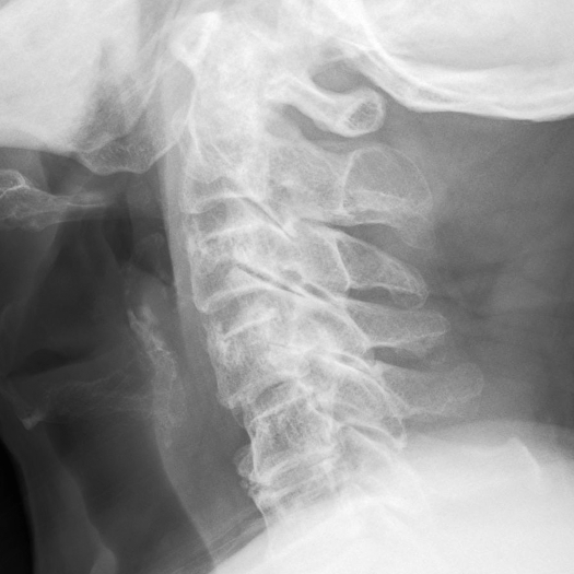
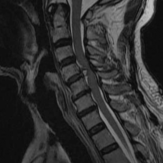
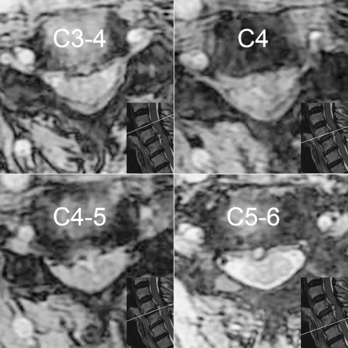
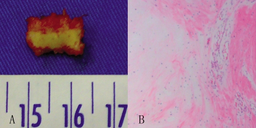
Findings: This 75-year-old man presented with acute paraplegia due to severe compression of the spinal cord by herniation of the C4-C5 cervical disk. He underwent emergency diskectomy and anterior fusion. Postoperatively, his neurologic functions improved gradually.
Conclusions: Cervical disk herniation should be considered in the differential diagnosis of nontraumatic acute paraplegia. Pre-existing narrowed canal is an important predisposing factor and excessive neck movements are believed to be triggering factors. Immediate early decompressive surgery is recommended to avoid irreversible progression of neurologic deficit.
Figures
References
-
- Kyriakides AE, Lalam RK, El Masry WS. Acute spontaneous spinal subdural hematoma presenting as paraplegia: a rare case. Spine. (Phila Pa 1976) 2007;32(21):E619–E622. - PubMed
-
- Rathore MF, Gill ZA, Malik AA, Farooq F. Acute flaccid paraplegia: a rare complication of meningococcal meningitis. Spinal Cord. 2008;46(4):314–316. - PubMed
-
- Huang MK, Chen CC, Wang TL, Lee YK, Su YC. Simultaneous bilateral femoral arterial emboli presenting with acute paraplegia: an uncommon case. Am J Emerg Med. 2009;27(6):753.e3–4. - PubMed
-
- Lourie H, Shende MC, Stewart DH., Jr The syndrome of central cervical soft disk herniation. JAMA. 1973;226(3):302–305. - PubMed
-
- Ueyama T, Tamaki N, Kondoh T, Miyamoto H, Akiyama H, Nagashima T. Non-traumatic acute paraplegia associated with cervical disk herniation: a case report. Surg Neurol. 1999;52(2):204–206; discussion 206–207. - PubMed
Publication types
MeSH terms
LinkOut - more resources
Full Text Sources
Medical
Miscellaneous