. 2010 Dec 1;132(47):16893-9.
doi: 10.1021/ja106553e. Epub 2010 Nov 9.

Biocompatible copper(I) catalysts for in vivo imaging of glycans

Affiliations

Biocompatible copper(I) catalysts for in vivo imaging of glycans

David Soriano Del Amo et al. J Am Chem Soc. .

Abstract

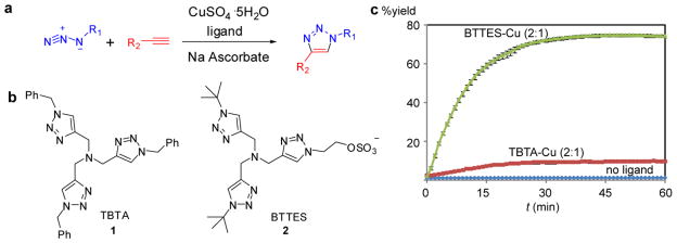
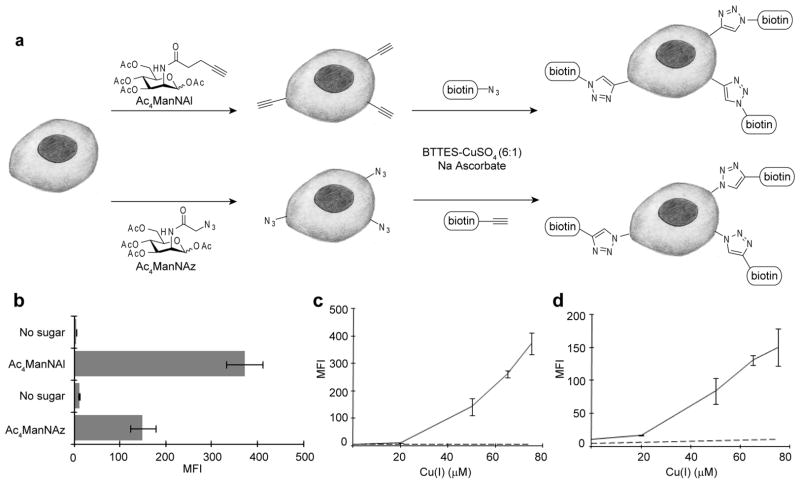
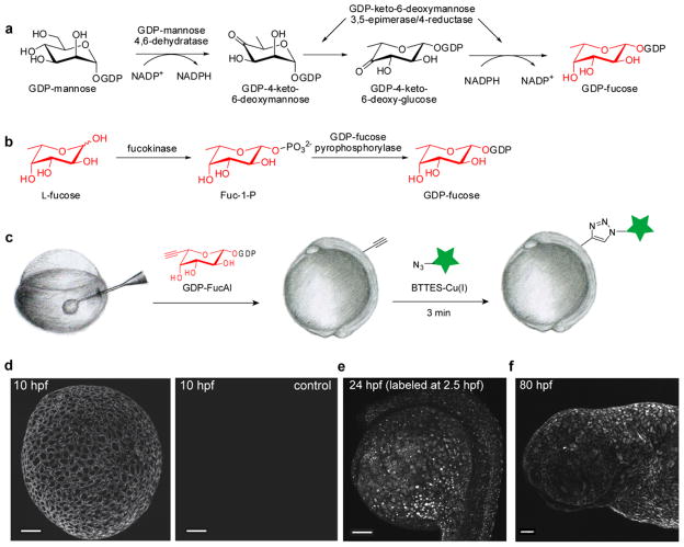
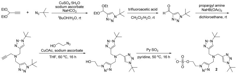
The Cu(I)-catalyzed azide-alkyne cycloaddition (CuAAC) is the standard method for bioorthogonal conjugation. However, current Cu(I) catalyst formulations are toxic, hindering their use in living systems. Here we report that BTTES, a tris(triazolylmethyl)amine-based ligand for Cu(I), promotes the cycloaddition reaction rapidly in living systems without apparent toxicity. This catalyst allows, for the first time, noninvasive imaging of fucosylated glycans during zebrafish early embryogenesis. We microinjected embryos with alkyne-bearing GDP-fucose at the one-cell stage and detected the metabolically incorporated unnatural sugars using the biocompatible click chemistry. Labeled glycans could be imaged in the enveloping layer of zebrafish embryos between blastula and early larval stages. This new method paves the way for rapid, noninvasive imaging of biomolecules in living organisms.


Figures

Figure 1
Figure 1
Copper(I)-catalyzed azide-alkyne cycloaddtion (CuAAC) is accelerated by Cu(I)-stabilizing ligands. (a) CuAAC of azides and terminal alkynes to form 1,4-disubstituted 1,2,3-triazoles. (b) Structures of CuAAC-accelerating ligands. (c) Conversion–time profiles of CuAAC in the presence/absence of accelerating ligands. Reaction conditions: propargyl alcohol (50 μM), 3-azido-7-hydroxy-coumarin (100 μM), CuSO4 (75 μM), 0.1 M potassium phosphate buffer (pH 7.0)/DMSO 95:5, sodium ascorbate (2.5 μM), room temperature. Error bars represent the standard deviation of three replicate experiments.
Figure 2
Figure 2
Detection of glycoconjugates on the surface of live cells via biocompatible CuAAC. (a) Schematic representation of metabolic labeling and detection of cell-surface sialic acids using Ac4ManNAl and Ac4ManNAz and BTTES-Cu(I)-catalyzed click chemistry. (b, c, d) Flow cytometry data of cell surface labeling experiments described in (a) using LNCaP cells. (b) Cells were treated with biotin-azide or biotin-alkyne (100 μM) in the presence of the BTTES-Cu(I) catalyst ([Cu] = 75μM) for 1 or 2.5 min respectively before probing with streptavidin-Alexa Fluor 488 conjugates. In all cases, cells cultured in the absence of sugar displayed mean fluorescence intensity (MFI, arbitrary units) values < 15. (c) Cells were labeled with biotin-azide (100 μM) for 1 min in the presence of 25–75 μM Cu(I). (d) Cells were labeled with biotin-alkyne (100 μM) for 2.5 min in the presence of 25–75 μM Cu(I). Error bars represent the standard deviation of three replicate experiments. Solid line, + Ac4ManNAl (c) or Ac4ManNAz (d); dashed line, no sugar.
Figure 3
Figure 3
Analysis of CuAAC kinetics on the surface of CHO cells by flow cytometry. CHO cells were incubated for 3 days in untreated medium or medium supplemented with 50 μM Ac4ManNAz, followed by labeling with 100 μM biotin-alkyne in the presence of BTTES-Cu(I) catalyst ([Cu] = 75 μM). The reaction was quenched at various time points with BCS and probed with Alexa Fluor 488-streptavidin conjugates. Error bars represent the standard deviation of three replicate experiments.
Figure 4
Figure 4
BTTES-Cu(I)-catalyzed click chemistry has no long term perturbation to Jurkat and HEK cells. (a) Cell growth curve after click chemistry treatment. (b) Cell viability measured using an alamarBlue assay (Invitrogen). Error bars represent the standard derivation for three replicates.
Figure 5
Figure 5
In vivo imaging of fucosylated glycans during early zebrafish embryogenesis via BTTES-Cu(I)-catalyzed click chemistry. (a) GDP-Fuc de novo biosynthetic pathway. (b) GDP-Fuc salvage pathway. (c) Microinjection combined with the BTTES-Cu(I)-catalyzed click chemistry enables the labeling of fucosylated glycans in zebrafish embryos. Zebrafish embryos were microinjected with a single dose of GDP-FucAl and allowed to develop to 10 hpf (d), 2.5 hpf (e) and 80 hpf (f). The embryos were then reacted with Alexa Fluor 488-azide catalyzed by BTTES-Cu(I) and imaged using confocal microscopy. The maximum intensity z-projection fluorescence image for e was acquired at 24 hpf. Scale bar: 100 μm.
Scheme 1
Scheme 1
The synthesis of BTTES.

References

    1. Kolb HC, Finn MG, Sharpless KB. Angew Chem Int Ed. 2001;40:2004–2021. - PubMed
    1. Baskin JM, Bertozzi CR. Qsar Comb Sci. 2007;26:1211–1219.
    1. Wang L, Schultz PG. Angew Chem Int Ed. 2004;44:34–66. - PubMed
    1. Laughlin ST, Agard NJ, Baskin JM, Carrico IS, Chang PV, Ganguli AS, Hangauer MJ, Lo A, Prescher JA, Bertozzi CR. Methods Enzymol. 2006;415:230–250. - PubMed
    1. Dieterich DC, Lee JJ, Link AJ, Graumann J, Tirrell DA, Schuman EM. Nat Protc. 2007;2:532–540. - PubMed

Publication types

LinkOut - more resources