Review
. 2010 Dec:1214:156-67.
doi: 10.1111/j.1749-6632.2010.05819.x. Epub 2010 Nov 9.

Genetic regulation of human brain development: lessons from Mendelian diseases

Affiliations
Review

Genetic regulation of human brain development: lessons from Mendelian diseases

Tracy J Dixon-Salazar et al. Ann N Y Acad Sci. 2010 Dec.

Abstract

One of the fundamental goals in human genetics is to link gene function to phenotype, yet the function of the majority of the genes in the human body is still poorly understood. This is especially true for the developing human brain. The study of human phenotypes that result from inherited, mutated alleles is the most direct evidence for the requirement of a gene in human physiology. Thus, the study of Mendelian central nervous system (CNS) diseases can be an extremely powerful approach to elucidate such phenotypic/genotypic links and to increase our understanding of the key components required for development of the human brain. In this review, we highlight examples of how the study of inherited neurodevelopmental disorders contributes to our knowledge of both the "normal" and diseased human brain, as well as elaborate on the future of this type of research. Mendelian disease research has been, and will continue to be, key to understanding the molecular mechanisms that underlie human brain function, and will ultimately form a basis for the design of intelligent, mechanism-specific treatments for nervous system disorders.


Conflict of interest statement

Conflicts of interest

The authors declare no conflicts of interest.

Figures

Figure 1
Figure 1
Overview of early cortical neuron development. Cortical neurons are born and undergo fate determination in the ventricular zone (VZ) and subventricular zone of the developing cortex. These neurons then migrate through the intermediate zone (IZ) and into the cortical plate (CP), where they come to reside in their specific cortical layer. Maturing neurons then send out axons, which grow outward until they reach their designated target. This process is repeated with each newly born neuron. Ultimately, neurons form many synaptic connections to create functional neural circuits. Mendelian disorders can occur at any of these developmental stages, and specific examples include primary microcephaly (MCPH), classical lissencephaly (LIS), horizontal gaze palsy with progressive scoliosis (HGPPS), and primary epilepsy.
Figure 2
Figure 2
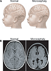
Anatomical features of primary microcephaly (MCPH). (A) Children with MCPH have a reduced head circumference beginning at birth and a small, but architecturally normal, brain as compared to a healthy control. Figure reprinted with permission. (B) MRI scans of a healthy child and a child with microcephaly. The microcephalic brain shows reduced brain volume, with the largest volume loss seen in the cortical areas.
Figure 3
Figure 3
Genes that cause MCPH localize to the centrosome during the cell cycle and play a role in neurogenesis. During neurogenic proliferation, progenitors first undergo symmetric cell division where two daughter cells of progenitor fate are produced. This is followed by asymmetric cell division in which one daughter cell follows a progenitor cell fate and the other follows a neural fate.,– Committed neurons then migrate to the cortical plate (CP). The cell cycle is critically regulated by the mitotic machinery,,, and each of the five MCPH genes (MCPH1, APSM, STIL, CENPJ, and CKD5RAP2) colocalize with the mitotic apparatus during at least some part of the cell cycle. Genetic studies reveal that MCPH is primarily a disorder of cortical neurogenesis and not a disorder of migration, cell death, or cell growth and provide insight into the genes that regulate brain size in humans.
Figure 4
Figure 4
Anatomical features of classical lissencephaly (LIS). Children with LIS, also known as smooth brain syndrome, lack folds and ridges in the outer cortical layers and typically have a smaller head size as compared to healthy controls (wm, white matter). Figure reprinted with permission.
Figure 5
Figure 5
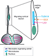
Genes that cause LIS localize to the leading edge of cortical neurons and play a role in neuron migration. Committed neurons migrate along radial glia to reach their specified layer in the developing cortical plate. Neuron migration involves a strictly regulated process of cytoskeletal rearrangements. Each of the LIS-causing genes (DCX, LIS1,TUBA1A) act as key regulators of microtubules. Genetic studies reveal that LIS is primarily a disorder of neuron migration and provide mechanistic information about the genetic regulation of cortical development in humans.
Figure 6
Figure 6
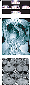
Anatomical features of horizontal gaze palsy with progressive scoliosis (HGPPS). (A) Photo of a patient with HGPPS showing lack of horizontal eye movements when attempting to look to either side. Upward and downward eye movements are normal. (B) MRI of the spine of a patient with HGPPS shows profound scoliosis. (C) MRI scans from a normal brain (a,b) and a patient with HGPPS (c,d). Patients with HGPPS display pons hypoplasia (boxed region in a,b); absent protrusions of the abducens nuclei (arrowheads in a,b); and flat, butterfly-like appearance of the medulla (open arrow in c,d). Figures reprinted with permission.
Figure 7
Figure 7
Many of the genes that cause primary epilepsy are cell surface receptors that act to regulate neural circuit formation and network signaling. During circuit formation, neurons from different brain areas form stereotypic synaptic connections. Circuit formation is initially neuronal activity independent, but as signaling receptors are expressed at the cell surface, neuronal activity begins to shape circuit structure and function. Thirteen of the identified genes that cause primary epilepsy fall into the voltage-gated or ligand-gated ion channel class and are expressed in the axon or at the synapse in various neuronal types. Studies reveal that epilepsy can result from a primary defect in ion channel function and also provide evidence that certain types of epilepsy result from defects in circuit formation and function in the developing neocortex.

References

    1. Drachman DA. Do we have brain to spare? Neurology. 2005;64:2004–2005. - PubMed
    1. International Human Genome Sequencing Consortium. Finishing the euchromatic sequence of the human genome. Nature. 2004;431:931–945. - PubMed
    1. Peltonen L, et al. Lessons from studying monogenic disease for common disease. Hum. Mol. Genet. 2006;15(Spec. No. 1):R67–R74. - PubMed
    1. Engel J, Pedley TA. Epilepsy: A Comprehensive Textbook. Philadelphia: Lippincott-Raven; 1998.
    1. Engle EC. Human genetic disorders of axon guidance. Cold Spring Harb. Perspect. Biol. 2010;2:a001784–a001802. - PMC - PubMed

Publication types

MeSH terms